. 2017 Dec 4;14(1):238.
doi: 10.1186/s12974-017-1015-2.

Neuronal CCL2 expression drives inflammatory monocyte infiltration into the brain during acute virus infection

Affiliations

Neuronal CCL2 expression drives inflammatory monocyte infiltration into the brain during acute virus infection

Charles L Howe et al. J Neuroinflammation. .

Abstract

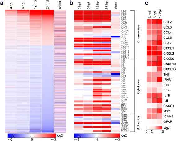
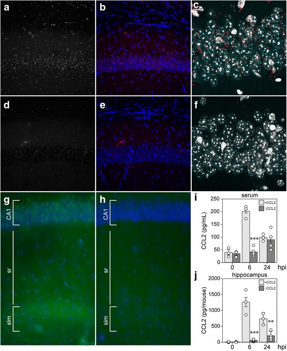
Background: Viral encephalitis is a dangerous compromise between the need to robustly clear pathogen from the brain and the need to protect neurons from bystander injury. Theiler's murine encephalomyelitis virus (TMEV) infection of C57Bl/6 mice is a model of viral encephalitis in which the compromise results in hippocampal damage and permanent neurological sequelae. We previously identified brain-infiltrating inflammatory monocytes as the primary driver of this hippocampal pathology, but the mechanisms involved in recruiting these cells to the brain were unclear.

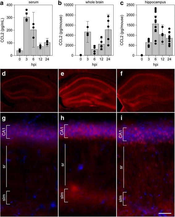
Methods: Chemokine expression levels in the hippocampus were assessed by microarray, ELISA, RT-PCR, and immunofluorescence. Monocyte infiltration during acute TMEV infection was measured by flow cytometry. CCL2 levels were manipulated by immunodepletion and by specific removal from neurons in mice generated by crossing a line expressing the Cre recombinase behind the synapsin promoter to animals with floxed CCL2.

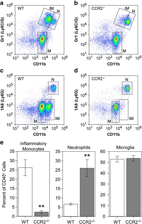
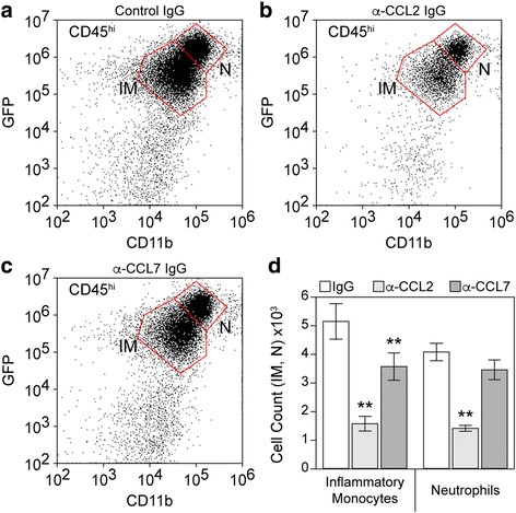
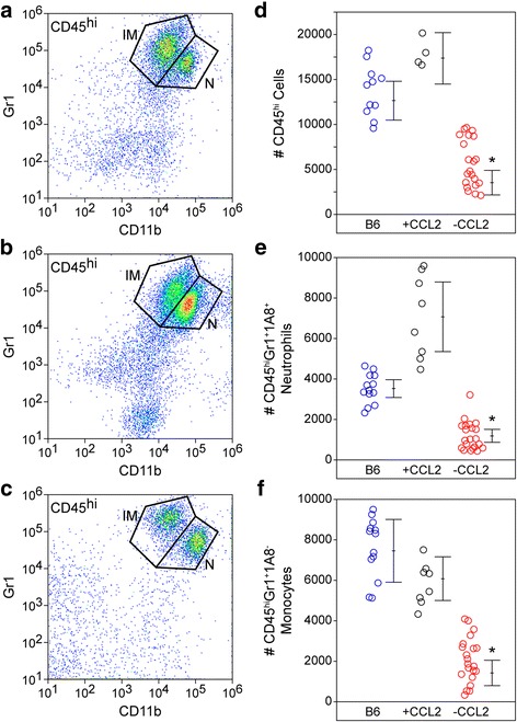
Results: Inoculation of the brain with TMEV induced hippocampal production of the proinflammatory chemokine CCL2 that peaked at 6 h postinfection, whereas inoculation with UV-inactivated TMEV did not elicit this response. Immunofluorescence revealed that hippocampal neurons expressed high levels of CCL2 at this timepoint. Genetic deletion of CCR2 and systemic immunodepletion of CCL2 abrogated or blunted the infiltration of inflammatory monocytes into the brain during acute infection. Specific genetic deletion of CCL2 from neurons reduced serum and hippocampal CCL2 levels and inhibited inflammatory monocyte infiltration into the brain.

Conclusions: We conclude that intracranial inoculation with infectious TMEV rapidly induces the expression of CCL2 in neurons, and this cellular source is necessary for CCR2-dependent infiltration of inflammatory monocytes into the brain during the most acute stage of encephalitis. These findings highlight a unique role for neuronal production of chemokines in the initiation of leukocytic infiltration into the infected central nervous system.

Keywords: CCL2; CCR2; Encephalitis; Hippocampus; Inflammatory monocyte; Theiler’s murine encephalomyelitis virus.


Conflict of interest statement

Ethics approval

All animal experiments were performed according to the National Institutes of Health guidelines and were approved by the Mayo Clinic Institutional Animal Care and Use Committee (Animal Welfare Assurance number A3291-01).

Consent for publication

Not applicable.

Competing interests

The authors declare that they have no competing interests.

Publisher’s Note

Springer Nature remains neutral with regard to jurisdictional claims in published maps and institutional affiliations.

Figures

Fig. 1
Fig. 1
Transcriptional program initiated in the hippocampus by acute infection. Hippocampal tissue was collected for RNA purification at early timepoints following intracranial inoculation of adult female C57Bl/6 mice with 2 × 105 PFU of the Daniel’s strain of TMEV. Transcriptional changes were analyzed using Illumina MouseWG-6 v2.0 beadchips. Fold change relative to uninfected control for 16,900 genes present on the array was calculated, converted to log2, and heatmapped using Gitools. Sham controls were injected intracranially with culture media used for the virus preparation, and RNA was collected at 24 h postinfection (hpi). Expression data for all conditions except sham are the means from six individual mice analyzed in two separate experiments (three mice per experiment). Three sham mice were analyzed in the second experiment. a Heatmap showing pattern of transcriptional changes at 3, 6, 12, and 24 hpi relative to uninfected controls. The discontinuity in expression coding is the result of removing from analysis all genes with changes between − 1.2-fold and + 1.2-fold at 24 hpi, leaving 6764 genes in the heatmap. Color-coded heatmaps range from greater than eightfold downregulated (log2 < − 3.0; blue) to greater than eightfold upregulated (log2 > + 3.0; red). b The same expression data remapped to highlight chemokines, cytokines, and adhesion factors. In contrast to (a), values are continuous between negative eightfold and positive eightfold. Note that the color range was selected to reveal weaker changes in some genes, resulting in saturation at all timepoints for highly upregulated genes such as CCL2. c A further subset of highly upregulated and notable genes was validated by RT-PCR and the results heatmapped on a scale from 0 to 1000-fold (log2 = + 10) to reveal large expression changes over the first 12 h
Fig. 2
Fig. 2
CCL2 levels in serum, whole brain, and hippocampus during acute infection. Serum (a), whole brain (b), and microdissected hippocampus (c) were collected at 0, 3, 6, 12, and 24 h following intracranial inoculation with TMEV. Absolute levels of CCL2 were measured by ELISA. Each dot represents an individual animal. The bar graph shows mean ± 95% confidence intervals from aggregate data. A minimum of three mice were used per tissue per timepoint, and the hippocampal samples were collected in a separate experiment from the whole brain samples. All results were analyzed by one-way ANOVA. CCL2 was induced in serum, whole brain, and hippocampus (P < 0.001 for each factor in each tissue compared to 0 hpi). Brain sections collected at 0 (d, g), 6 (e, h), and 24 hpi (f, i) were immunostained to reveal CCL2 expression. In the hippocampus, there was weak, diffuse signal in uninfected mice (d, g) that increased markedly at 6 hpi (e, h). By 24 hpi, the signal intensity had decreased and exhibited a more punctate pattern (f, i). Scale bar in i is 50 μm and refers to g–i. CA1 cornu ammonis 1 formation, sr stratum radiatum, slm stratum lacunosum moleculare. Immunostaining is representative of more than three mice per timepoint in more than three separate experiments
Fig. 3
Fig. 3
Inflammatory monocyte infiltration during acute infection requires CCR2. Brain-infiltrating leukocytes were collected from C57Bl/6 mice (WT; a, c) and CCR2−/− mice (b, d) at 18 hpi. Flow plots in (ad) show cells in a CD45+ parent gate. The number of inflammatory monocytes (IM; CD45hiCD11b+Gr1++1A8), neutrophils (N; CD45hiCD11b++Gr1+1A8+), and microglia (M; CD45midCD11bmidGr11A8) was measured by flow cytometry. The percent of inflammatory monocytes, neutrophils, and microglia in the CD45+ population is shown as mean ± 95%CI calculated from nine mice per genotype in three separate experiments (3 × 3) (e). Data were analyzed by Dunnett’s method. No difference in microglia was observed, but neutrophils were significantly increased in CCR2−/− mice (P = 0.0012 vs WT). Notably, the absence of CCR2 robustly inhibited inflammatory monocyte infiltration (P = 0.0004 vs WT). **P < 0.001
Fig. 4
Fig. 4
Inflammatory monocyte infiltration during acute infection is driven predominantly by CCL2. LysM:GFP mice received 20 μg goat IgG (a), 20 μg goat anti-CCL2 IgG (b), or 20 μg goat anti-CCL7 IgG (c) by intraperitoneal injection at − 12, 0, and + 12 h relative to time of infection. Brain-infiltrating leukocytes were collected at 24 hpi. The flow plots in (ac) show cells in a CD45hi parent gate. The number of inflammatory monocytes (IM: CD45hiCD11b+GFPmid) and neutrophils (N: CD45hiCD11b++GFPhi) were counted; values are shown as mean ± 95% CI calculated from six mice per treatment condition in two separate experiments (3 × 2) (d). Data were analyzed by one-way ANOVA with Dunnett’s method for pairwise comparison to control. Inflammatory monocytes were significantly reduced in both anti-chemokine groups compared to control IgG; neutrophils were significantly reduced only in the anti-CCL2 IgG group; **P < 0.001
Fig. 5
Fig. 5
Neurons are the primary source of CCL2 at 6 hpi. Ccl2-RFPfl/fl reporter mice (ac) and Syn-Cre x Ccl2-RFPfl/fl neuron-specific CCL2-deficient mice (df) were intracranially inoculated with TMEV and brain sections were collected at 6 hpi for analysis of RFP expression. Single-channel RFP (a, d) and two-channel RFP (red) and DAPI (blue) (b, e) microscopy revealed that the reporter signal present in neurons at 6 hpi was specifically deleted in the Syn-Cre x Ccl2-RFPfl/fl mice. Higher power imaging of the CA1 pyramidal neuron layer verified the presence of reporter signal (red) in these neurons (marked by DAPI; white) in Ccl2-RFPfl/fl mice (c) and the almost complete absence of such signal in the conditional knockout (f). Immunostaining with anti-CCL2 antibody confirmed the presence of CCL2 (green) in CA1 neurons (marked by DAPI; blue) and in the stratum lacunosum moleculare at 6 hpi in Ccl2-RFPfl/fl mice (g) and the absence of CCL2 in the hippocampus of Syn-Cre x Ccl2-RFPfl/fl conditional knockout mice (h). Serum (i) and hippocampal (j) CCL2 levels were measured by ELISA at 0, 6, and 24 hpi in the CCL2 reporter mice (+CCL2) and the neuron-specific CCL2-deficient mice (−CCL2). Each dot represents one animal; bar graphs represent mean ± 95%CI calculated from at least three mice per group per timepoint. Data were analyzed by two-way ANOVA with Tukey-Kramer pairwise analysis; statistical significance is only shown between genotypes at each timepoint; **P < 0.001; ***P < 0.0001. CA1 cornu ammonis 1 formation, sr stratum radiatum, slm stratum lacunosum moleculare. Fluorescence in af is representative of more than three mice in two separate experiments; immunostaining in gh is representative of two animals in each group
Fig. 6
Fig. 6
Loss of acute neuronal CCL2 production results in reduced inflammatory monocyte infiltration into the brain. Brain-infiltrating leukocytes were analyzed by flow cytometry in wildtype B6 mice (a), Ccl2-RFPfl/fl reporter mice (+CCL2; b), and Syn-Cre x Ccl2-RFPfl/fl neuron-specific CCL2-deficient mice (−CCL2; c) at 18 hpi. The flow plots in (ac) show Gr1- and CD11b-labeled cells in a CD45hi parent gate. The number of CD45hi cells (d), neutrophils (e), and inflammatory monocytes (f) were counted in the three groups (blue circles = B6; black circles = +CCL2; red circles = −CCL2). Each dot represents one animal; the line graph represents mean ± 95%CI calculated from at least two separate experiments. All cell types were significantly reduced in the neuron-specific CCL2-deficient mice but not in the parent reporter line. Data were analyzed by one-way ANOVA with Dunnett’s pairwise comparison; *P ≤ 0.0001

Similar articles

Cited by

References

    1. Shives KD, Tyler KL, Beckham JD. Molecular mechanisms of neuroinflammation and injury during acute viral encephalitis. J Neuroimmunol. 2017;308:102–111. doi: 10.1016/j.jneuroim.2017.03.006. - DOI - PubMed
    1. Buenz EJ, Rodriguez M, Howe CL. Disrupted spatial memory is a consequence of picornavirus infection. Neurobiol Dis. 2006;24:266–273. doi: 10.1016/j.nbd.2006.07.003. - DOI - PubMed
    1. DePaula-Silva AB, Hanak TJ, Libbey JE, Fujinami RS. Theiler’s murine encephalomyelitis virus infection of SJL/J and C57BL/6J mice: models for multiple sclerosis and epilepsy. J Neuroimmunol. 2017;308:30–42. doi: 10.1016/j.jneuroim.2017.02.012. - DOI - PMC - PubMed
    1. Howe CL, Lafrance-Corey RG, Sundsbak RS, Lafrance SJ. Inflammatory monocytes damage the hippocampus during acute picornavirus infection of the brain. J Neuroinflammation. 2012;9:50. doi: 10.1186/1742-2094-9-50. - DOI - PMC - PubMed
    1. Umpierre AD, Remigio GJ, Dahle EJ, Bradford K, Alex AB, Smith MD, West PJ, White HS, Wilcox KS. Impaired cognitive ability and anxiety-like behavior following acute seizures in the Theiler’s virus model of temporal lobe epilepsy. Neurobiol Dis. 2014;64:98–106. doi: 10.1016/j.nbd.2013.12.015. - DOI - PMC - PubMed

LinkOut - more resources