

SP-A2 contributes to miRNA-mediated sex differences in response to oxidative stress: pro-inflammatory, anti-apoptotic, and anti-oxidant pathways are involved
- PMID: 29202868
- PMCID: PMC5716385
- DOI: 10.1186/s13293-017-0158-2
SP-A2 contributes to miRNA-mediated sex differences in response to oxidative stress: pro-inflammatory, anti-apoptotic, and anti-oxidant pathways are involved
Abstract
Background: Human innate host defense molecules, surfactant protein A1 (SP-A1), and SP-A2 differentially affect the function and proteome of the alveolar macrophage (AM). We hypothesized that SP-A genes differentially regulate the AM miRNome.
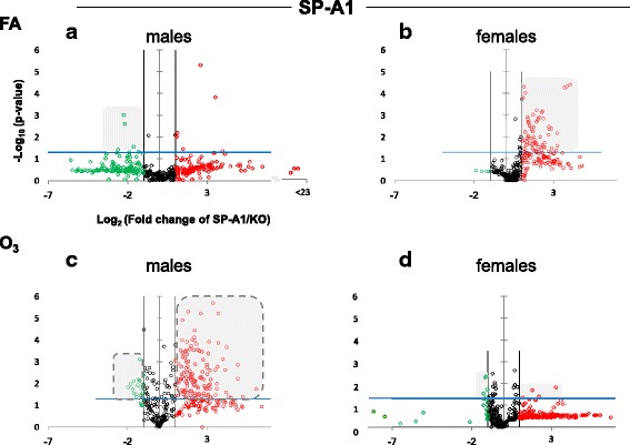
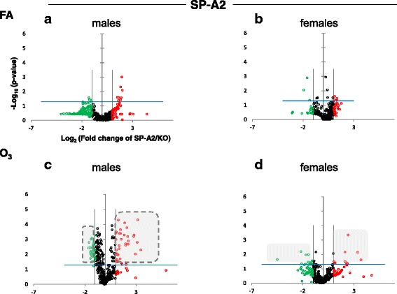
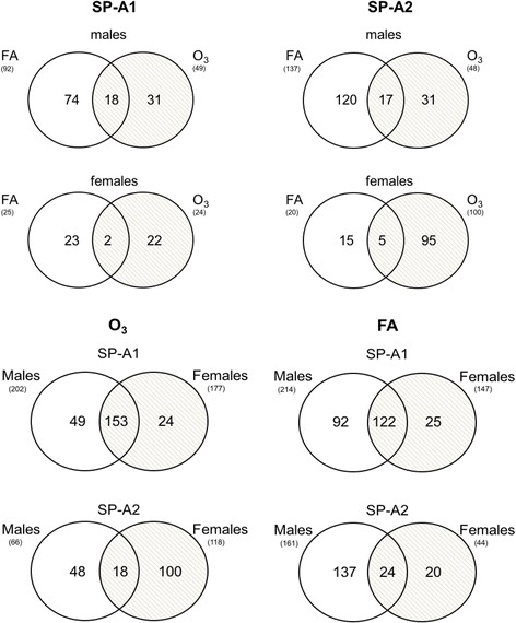
Methods: Humanized transgenic mice expressing SP-A1 and SP-A2 were subjected to O3-induced oxidative stress (OxS) or filtered air (FA), AMs were isolated, and miRNA levels were measured.
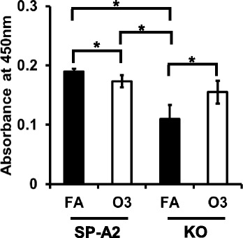
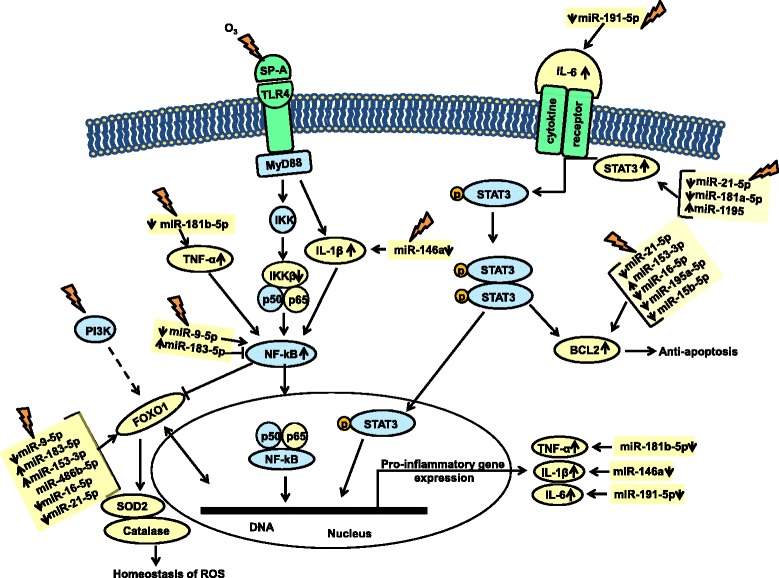
Results: In SP-A2 males, we found significant changes in miRNome in terms of sex and sex-OxS effects, with 11 miRNAs differentially expressed under OxS. Their mRNA targets included BCL2, CAT, FOXO1, IL6, NF-kB, SOD2, and STAT3. We followed the expression of these transcripts as well as key cytokines, and we found that (a) the STAT3 mRNA significantly increased at 4 h post OxS and returned to baseline at 18 h post OxS. (b) The anti-oxidant protein SOD2 level significantly increased, but the CAT level did not change after 4 h post OxS compared to control. (c) The anti-apoptotic BCL2 mRNA increased significantly (18 h post OxS), but the levels of the other transcripts were decreased. The presence of the SP-A2 gene had a protective role in apoptosis of AMs under OxS compared to mice lacking SP-A (knockout, KO). (d) Pro-inflammatory cytokine IL-6 protein levels were significantly increased in SP-A2 mice compared to KO (4 and 18 h post OxS), which signifies the role of SP-A2 in pro-inflammatory protein expression. (e) SOD2 and CAT mRNAs changed significantly in OxS indicating a plausible role of SP-A2 in the homeostasis of reactive oxygen species. (f) Gonadectomy of transgenic mice showed that sex hormones contribute to significant changes of the miRNome expression.
Conclusions: We conclude that SP-A2 influences the miRNA-mediated sex-specific differences in response to OxS. In males, these differences pertain to inflammatory, anti-apoptotic, and anti-oxidant pathways.
Keywords: IL-6; Ozone; STAT3; Sex differences; Surfactant protein A.
Conflict of interest statement
Ethics approval
All protocols used in this study were evaluated and approved by the Pennsylvania State University College of Medicine Institutional Animal Care and Use Committee and conformed to the guidelines of the National Institutes of Health on the care and use of laboratory animals.
Consent for publication
Not applicable
Competing interests
The authors declare that they have no competing interests.
Publisher’s Note
Springer Nature remains neutral with regard to jurisdictional claims in published maps and institutional affiliations.
Figures


Similar articles
-
Major Effect of Oxidative Stress on the Male, but Not Female, SP-A1 Type II Cell miRNome.Front Immunol. 2019 Jul 10;10:1514. doi: 10.3389/fimmu.2019.01514. eCollection 2019. Front Immunol. 2019. PMID: 31354704 Free PMC article.
-
Differential Impact of Co-expressed SP-A1/SP-A2 Protein on AM miRNome; Sex Differences.Front Immunol. 2019 Aug 16;10:1960. doi: 10.3389/fimmu.2019.01960. eCollection 2019. Front Immunol. 2019. PMID: 31475015 Free PMC article.
-
The Lung Alveolar Cell (LAC) miRNome and Gene Expression Profile of the SP-A-KO Mice After Infection With and Without Rescue With Human Surfactant Protein-A2 (1A0).Front Immunol. 2022 Jul 1;13:854434. doi: 10.3389/fimmu.2022.854434. eCollection 2022. Front Immunol. 2022. PMID: 35844510 Free PMC article.
-
Human Surfactant Protein SP-A1 and SP-A2 Variants Differentially Affect the Alveolar Microenvironment, Surfactant Structure, Regulation and Function of the Alveolar Macrophage, and Animal and Human Survival Under Various Conditions.Front Immunol. 2021 Aug 17;12:681639. doi: 10.3389/fimmu.2021.681639. eCollection 2021. Front Immunol. 2021. PMID: 34484180 Free PMC article. Review.
-
Genetic complexity of the human innate host defense molecules, surfactant protein A1 (SP-A1) and SP-A2--impact on function.Crit Rev Eukaryot Gene Expr. 2009;19(2):125-37. doi: 10.1615/critreveukargeneexpr.v19.i2.30. Crit Rev Eukaryot Gene Expr. 2009. PMID: 19392648 Free PMC article. Review.
Cited by
-
Major Effect of Oxidative Stress on the Male, but Not Female, SP-A1 Type II Cell miRNome.Front Immunol. 2019 Jul 10;10:1514. doi: 10.3389/fimmu.2019.01514. eCollection 2019. Front Immunol. 2019. PMID: 31354704 Free PMC article.
-
Profile of Rat Adrenal microRNAs Induced by Gonadectomy and Testosterone or Estradiol Replacement.Int J Mol Sci. 2025 May 9;26(10):4543. doi: 10.3390/ijms26104543. Int J Mol Sci. 2025. PMID: 40429687 Free PMC article.
-
The importance of translational science within the respiratory field.Breathe (Sheff). 2024 Mar;20(1):230183. doi: 10.1183/20734735.0183-2023. Epub 2024 May 14. Breathe (Sheff). 2024. PMID: 38746906 Free PMC article.
-
Can Prophylactic High Flow of Humidified and Warmed Filtered Air Improve Survival from Bacterial Pneumonia and SARS-CoV-2 in Elderly Individuals? The Role of Surfactant Protein A.Antioxidants (Basel). 2021 Apr 22;10(5):640. doi: 10.3390/antiox10050640. Antioxidants (Basel). 2021. PMID: 33922049 Free PMC article.
-
Survival of Surfactant Protein-A1 and SP-A2 Transgenic Mice After Klebsiella pneumoniae Infection, Exhibits Sex-, Gene-, and Variant Specific Differences; Treatment With Surfactant Protein Improves Survival.Front Immunol. 2018 Oct 16;9:2404. doi: 10.3389/fimmu.2018.02404. eCollection 2018. Front Immunol. 2018. PMID: 30459763 Free PMC article.
References
-
- Kremlev SG, Phelps DS. Effect of SP-A and surfactant lipids on expression of cell surface markers in the THP-1 monocytic cell line. Am J Phys. 1997;272:L1070–L1077. - PubMed
MeSH terms
Substances
Grants and funding
LinkOut - more resources
Full Text Sources
Other Literature Sources
Molecular Biology Databases
Research Materials
Miscellaneous
