. 2018 Jan 26;293(4):1192-1202.
doi: 10.1074/jbc.M117.797290. Epub 2017 Dec 4.

Crystal and solution structures of human protein-disulfide isomerase-like protein of the testis (PDILT) provide insight into its chaperone activity

Affiliations

Crystal and solution structures of human protein-disulfide isomerase-like protein of the testis (PDILT) provide insight into its chaperone activity

Huanhuan Li et al. J Biol Chem. .

Abstract

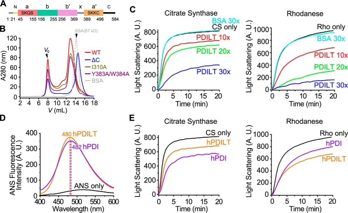
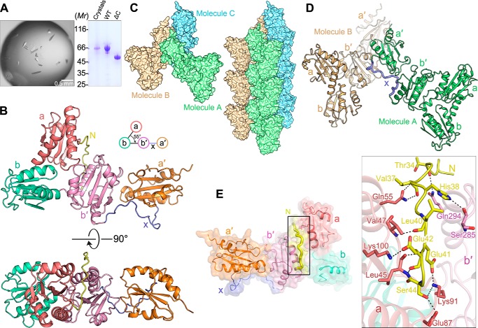
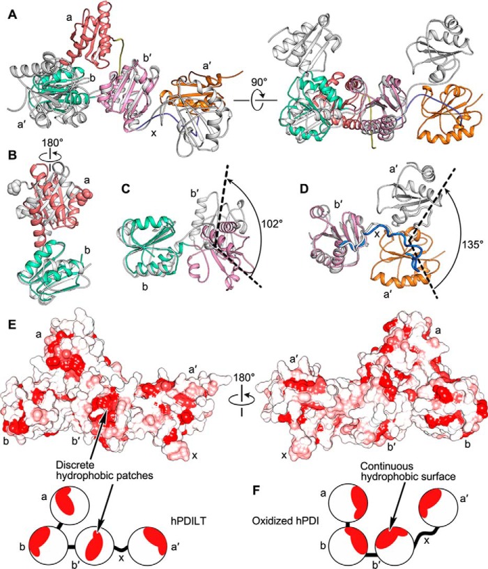
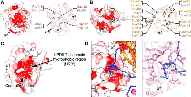
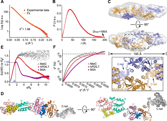
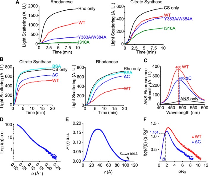
Protein-disulfide isomerase-like protein of the testis (PDILT), a member of the protein-disulfide isomerase family, is a chaperone essential for the folding of spermatogenesis-specific proteins in male postmeiotic germ cells. However, the structural mechanisms that regulate the chaperone function of PDILTs are unknown. Here, we report the structures of human PDILT (hPDILT) determined by X-ray crystallography to 2.4 Å resolution and small-angle X-ray scattering (SAXS). Distinct from previously reported U-like structures of related PDI family proteins, our structures revealed that hPDILT folds into a compact L-like structure in crystals and into an extended chain-like structure in solution. The hydrophobic regions and the hydrophobic pockets in hPDILT, which are important for substrate recognition, were clearly delineated in the crystal structure. Moreover, our results of the SAXS analysis and of structure-based substitutions and truncations indicated that the C-terminal tail in hPDILT is required for suppression of aggregation of denatured proteins, suggesting that the tail is crucial for the chaperone activity of PDILT. Taken together, our findings have identified the critical regions and conformational changes of PDILT that enable and control its activity. These results advance our understanding of the structural mechanisms involved in the chaperone activity of PDILT.

Keywords: X-ray crystallography; conformational change; molecular chaperone; protein structure; small-angle X-ray scattering (SAXS).


Conflict of interest statement

The authors declare that they have no conflicts of interest with the contents of this article

Figures

Figure 1.
Figure 1.
Chaperone activity of hPDILT. A, domain organization of hPDILT. The redox-inactive motifs are indicated in a and a′ domains. B, SEC profiles of WT, mutated, and C-terminally truncated hPDILT proteins. C, denatured and reduced citrate synthase (CS) (10 μm) or rhodanese (Rho) (22.5 μm) was 50-fold (for CS) or 100-fold (for Rho) diluted in the absence or presence of hPDILT or BSA as indicated in a molar ratio at 25 °C. A.U., arbitrary units. D, ANS fluorescence spectra of hPDILT and hPDI. 50 μm ANS was incubated without or with 5 μm hPDI and hPDILT for 20 min at 25 °C. ANS emission spectra were determined with excitation at 370 nm. E, chaperone activities of hPDILT and hPDI on CS or Rho were monitored as described in C. The molar ratio of hPDILT or hPDI to substrates is 10. A.U., arbitrary units.
Figure 2.
Figure 2.
Crystal structure of hPDILT. A, to test whether the C-terminal tail (Ile-497–Val-580) of hPDILT was degraded during crystallization, hPDILT crystals were washed with precipitant and dissolved in loading buffer for SDS-PAGE analysis. The band shown at 66 kDa suggests that the full-length hPDILT was present in the crystals. B, schematic diagrams of the crystal structure of hPDILT from the front view (upper panel) and the top view (lower panel). Color-coding of each domain is identical to that in Fig. 1A. C, crystal-packing reveals interactions between hPDILT molecules. D, x-linker of one hPDILT molecule A binds to the b′ domain of another symmetric molecule B. E, interactions between the N-terminal loop (Ser-32–Ser-44) and the thioredoxin-like a and b′ domains. Schematic and surface representation of hPDILT structure is colored as B. The N-terminal loop is highlighted in bold and yellow. Residues involved in the interaction are shown in stick representation on the right in the close-up view.
Figure 3.
Figure 3.
Differences between the structures of hPDILT and hPDI. A, superposition of hPDILT (colored as Fig. 2B) and oxidized hPDI (PDB code 4EL1, in gray) structures based on the b-b′ domains. B and C, comparison of both a-b and b-b′ domains of hPDILT and oxidized hPDI based on the b domains. The start residues of two a domains are shown in sphere representation. D, superimposition of b′–x-a′ domains of hPDILT and oxidized hPDI based on the b′ domains. E, hydrophobic surface representation of hPDILT, colored from hydrophobic (red) to hydrophilic (white), according to the normalized consensus hydrophobicity scale of the surface-exposed residues by UCSF Chimera (45). The lower panel shows the schematic diagram. F, schematic diagram of oxidized hPDI (PDB code 4EL1).
Figure 4.
Figure 4.
Hydrophobic regions in the thioredoxin-like domains of hPDILT. A, HRA of hPDILT is shown on the left. The conserved hydrophobic residues in structures of the a domains of hPDILT and hPDI are shown on the right. hPDI is shown in gray (PDB code 4EKZ). B, HRA′ of hPDILT and the conserved hydrophobic residues in structures of a′ domains of hPDILT and hPDI. C, HRB′ of hPDILT. D, close-up views of the interactions between b′ domain and x-linker from another molecule. The residues involved in hydrophobic interactions are shown as sticks.
Figure 5.
Figure 5.
SAXS analysis of the hPDILT structure in solution. A, overlay of the experimental scattering profile with the back-calculated scattering profile from the CORAL model of hPDILT. B, pair distance distribution P(r) functions for hPDILT. C, superposition of low-resolution ab initio model and rigid body model. The ab initio model is shown as light-gray surface representation. Two molecules (A and B) are shown in different colors (orange and blue). The close-up view shows the dimeric interface. D, solution structure of hPDILT monomer. The thioredoxin-like domains in the rigid body model are colored as in Fig. 2B and the N-terminal loop, x-linker, and C-terminal tail are represented in spheres, shown in yellow, blue, and gray, respectively. E, superimposition of dimensionless Kratky plot representations of experimental data. The SAXS data of MetC was downloaded from Small Angle Scattering Biological Data Bank (code SASDBY5). F, Kratky-Debye plot of each data set is described in E. a.u., arbitrary units.
Figure 6.
Figure 6.
Chaperone activities and SAXS analysis of hPDILT C-tail truncation. A and B, chaperone activities of WT hPDILT, I310A, and Y383A/W384A mutants and the C-tail truncation (ΔC) on CS or Rho were monitored as described in Fig. 1C. The molar ratio of hPDILT proteins to substrates is 10. C, ANS fluorescence spectra of hPDILT and ΔC were monitored as described in Fig. 1D. A.U., arbitrary units. D, experimental scattering profile of ΔC hPDILT protein. E, pair distance distribution P(r) functions for ΔC hPDILT protein. F, superimposition of dimensionless Kratky plot representations of wildtype and ΔC hPDILT experimental data.

References

    1. Lipton S. A., Gu Z., and Nakamura T. (2007) Inflammatory mediators leading to protein misfolding and uncompetitive/fast off-rate drug therapy for neurodegenerative disorders. Int. Rev. Neurobiol. 82, 1–27 10.1016/S0074-7742(07)82001-0 - DOI - PubMed
    1. Labbadia J., and Morimoto R. I. (2015) The biology of proteostasis in aging and disease. Annu. Rev. Biochem. 84, 435–464 10.1146/annurev-biochem-060614-033955 - DOI - PMC - PubMed
    1. Sitia R., and Braakman I. (2003) Quality control in the endoplasmic reticulum protein factory. Nature 426, 891–894 10.1038/nature02262 - DOI - PubMed
    1. Wang L., Wang X., and Wang C.-C. (2015) Protein disulfide–isomerase, a folding catalyst and a redox-regulated chaperone. Free Radic. Biol. Med. 83, 305–313 10.1016/j.freeradbiomed.2015.02.007 - DOI - PubMed
    1. Kozlov G., Määttänen P., Thomas D. Y., and Gehring K. (2010) A structural overview of the PDI family of proteins. FEBS J. 277, 3924–3936 10.1111/j.1742-4658.2010.07793.x - DOI - PubMed

Publication types

Substances

LinkOut - more resources