. 2017 Dec 4;7(1):16837.
doi: 10.1038/s41598-017-17082-x.

Structures of PPARγ complexed with lobeglitazone and pioglitazone reveal key determinants for the recognition of antidiabetic drugs

Affiliations

Structures of PPARγ complexed with lobeglitazone and pioglitazone reveal key determinants for the recognition of antidiabetic drugs

Min A Lee et al. Sci Rep. .

Abstract

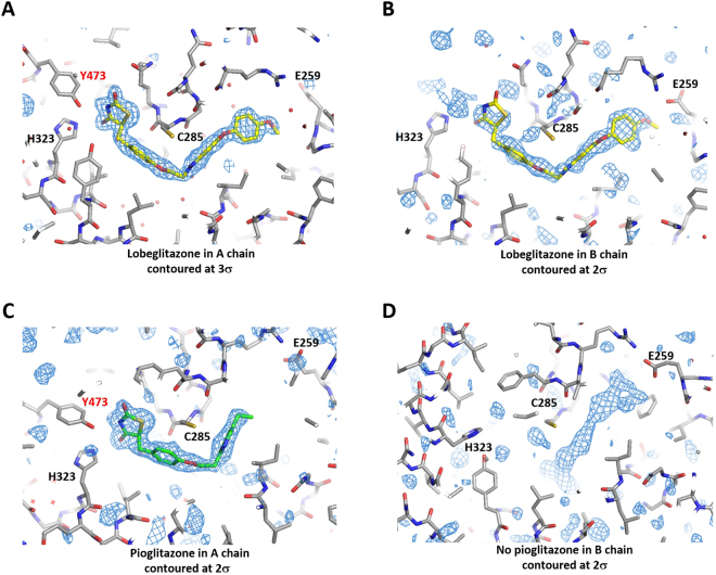
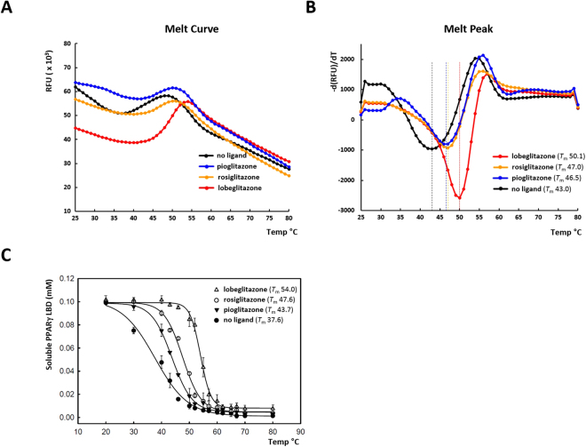
Peroxisome proliferator-activator receptor (PPAR) γ is a nuclear hormone receptor that regulates glucose homeostasis, lipid metabolism, and adipocyte function. PPARγ is a target for thiazolidinedione (TZD) class of drugs which are widely used for the treatment of type 2 diabetes. Recently, lobeglitazone was developed as a highly effective TZD with reduced side effects by Chong Kun Dang Pharmaceuticals. To identify the structural determinants for the high potency of lobeglitazone as a PPARγ agonist, we determined the crystal structures of the PPARγ ligand binding domain (LBD) in complex with lobeglitazone and pioglitazone at 1.7 and 1.8 Å resolutions, respectively. Comparison of ligand-bound PPARγ structures revealed that the binding modes of TZDs are well conserved. The TZD head group forms hydrogen bonds with the polar residues in the AF-2 pocket and helix 12, stabilizing the active conformation of the LBD. The unique p-methoxyphenoxy group of lobeglitazone makes additional hydrophobic contacts with the Ω-pocket. Docking analysis using the structures of TZD-bound PPARγ suggested that lobeglitazone displays 12 times higher affinity to PPARγ compared to rosiglitazone and pioglitazone. This structural difference correlates with the enhanced affinity and the low effective dose of lobeglitazone compared to the other TZDs.


Conflict of interest statement

The authors declare that they have no competing interests.

Figures

Figure 1
Figure 1
Overall structures of the PPARγ LBD complexed with pioglitazone and lobeglitazone. (A) Chemical structures of pioglitazone, rosiglitazone, and lobeglitazone. Structurally conserved parts of TZDs are shaded with light blue colors. (B) Monomeric structure of the PPARγ LBDs complexed with lobeglitazone. The C-terminal AF-2 helix (H12) is colored in red. The disordered Ω-loops are indicated with dashed lines. The bound ligands are shown as transparent spheres. (C) Monomeric structure of the PPARγ LBDs complexed with pioglitazone. (D) The PPARγ LBD crystallized as a homo-dimer composed of active and inactive forms in an asymmetric unit. (E) Structural superposition of A and B chains of the lobeglitazone-bound PPARγ LBD. (F) Structural comparison of the lobeglitazone- and pioglitazone-bound PPARγ LBDs.
Figure 2
Figure 2
Electron density maps of lobeglitazone and pioglitazone in the ligand-binding pockets of PPARγ. (A) 1.7 Å resolution F o-F c maps of lobeglitazone in A chain with the final model superimposed. (B) 1.7 Å resolution F o-F c maps of lobeglitazone in B chain. (C) 1.8 Å resolution F o-F c maps of pioglitazone in A chain. (D) 1.8 Å resolution F o-F c maps of the ligand-binding site in B chain of the pioglitazone-bound PPARγ LBD.
Figure 3
Figure 3
Structure of PPARγ – TZD interaction. (A) Surface representation of the Y-shaped ligand-binding cavity of PPARγ. The overall structure of the PPARγ LBD is shown in transparent ribbons. The bound lobeglitazone is shown in yellow spheres. (B) The surface representation of the ligand-binding pocket with lobeglitazone. Water molecules in the cavity are shown in small red spheres. (C) The surface representation of the ligand-binding pocket with pioglitazone. (D) The interaction of lobeglitazone and the PPARγ LBD was shown in 2-dimension using software Ligplot. The residues colored in green indicate the residues in the Ω-pocket. Water molecules are shown in cyan spheres. The hydrogen bonds are shown in dotted cyan lines. (E) The interaction of pioglitazone and the PPARγ LBD. (F) Structural comparison of the ligand binding modes for lobeglitazone, pioglitazone, and rosiglitazone. The structure of rosiglitazone was taken from the PDB id, 3DZY. The Tyr473 from H12 are colored in red.
Figure 4
Figure 4
Stabilization of the PPARγ LBD by ligand binding against heat denaturation. (A) The melting curves of the PPARγ LBD were monitored by differential scanning fluorimetry with SYPRO orange dye to examine the stabilization by ligand binding from thermal denaturation. (B) The reciprocal derivative plots of the melting curves. The dotted lines in the melt peak plots indicate T m values. (C) Stabilization of the PPARγ LBD against heat denaturation monitored by UV absorbance. The concentration of soluble PPARγ LBD was measured after heat denaturation from the starting protein concentration of 0.1 mM. The data points are means of three independent experiments and the error bars denote standard deviations.

Similar articles

Cited by

References

    1. DeFronzo RA, et al. Type 2 diabetes mellitus. Nat Rev Dis Primers. 2015;1:15019. doi: 10.1038/nrdp.2015.19. - DOI - PubMed
    1. Modi P. Diabetes beyond insulin: review of new drugs for treatment of diabetes mellitus. Curr Drug Discov Technol. 2007;4:39–47. doi: 10.2174/157016307781115476. - DOI - PubMed
    1. Han L, Shen WJ, Bittner S, Kraemer FB, Azhar S. PPARs: regulators of metabolism and as therapeutic targets in cardiovascular disease. Part II: PPAR-beta/delta and PPAR-gamma. Future Cardiol. 2017;13:279–296. doi: 10.2217/fca-2017-0019. - DOI - PMC - PubMed
    1. Kota BP, Huang TH, Roufogalis BD. An overview on biological mechanisms of PPARs. Pharmacol Res. 2005;51:85–94. doi: 10.1016/j.phrs.2004.07.012. - DOI - PubMed
    1. Nolte RT, et al. Ligand binding and co-activator assembly of the peroxisome proliferator-activated receptor-gamma. Nature. 1998;395:137–143. doi: 10.1038/25931. - DOI - PubMed

Publication types

MeSH terms