

Incidence of spinal epidural lipomatosis in patients with spinal stenosis
- PMID: 29203971
- PMCID: PMC5709284
- DOI: 10.1016/j.jor.2017.11.001
Incidence of spinal epidural lipomatosis in patients with spinal stenosis
Abstract
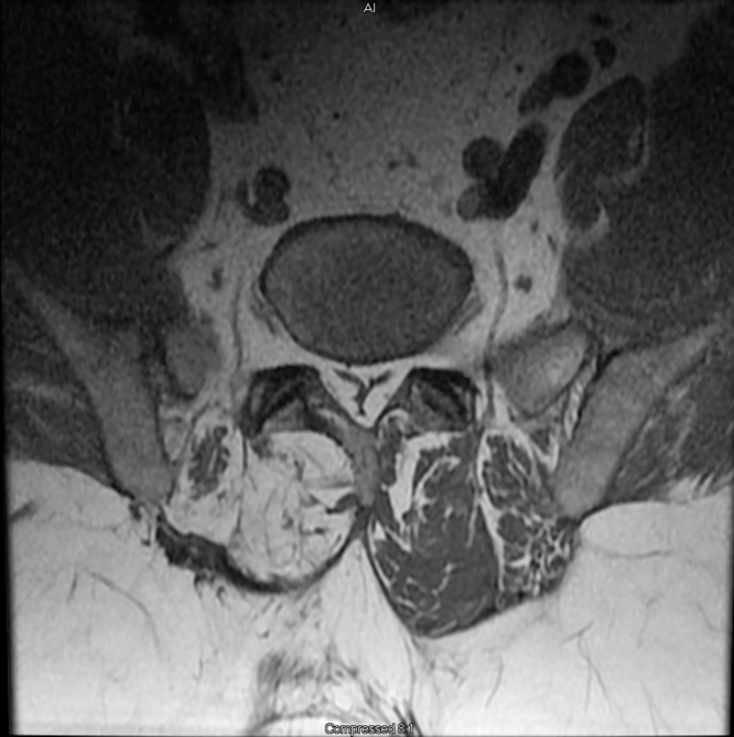
Inroduction: Spinal Epidural Lipomatosis (SEL) is believed to be a rare disorder. The incidence and prevalence of clinically symptomatic SEL in patients with spinal stenosis has never been reported in the literature. Our study aims to determine the prevalence, incidence, and associated risk factors of SEL in patients with the diagnosis of spinal stenosis.
Methods: This is a retrospective study. We reviewed the charts of 831 patients with the diagnosis of spinal stenosis over a 30 month period. All patients had spinal MRIs. Grading of SEL was performed using the Borré method.
Results: 52 patients (21 female and 31 male) had symptomatic moderate and severe SEL. We found a prevalence of 6.26% and an annual incidence of 2.5%. SEL was most commonly seen at L5-S1 level. 27% had received corticosteroids. All SEL patients were overweight and 79% were obese.
Conclusions: SEL is not uncommon in patients with spinal stenosis. SEL should be considered as a possible diagnosis in those with symptoms of spinal stenosis especially in those with associated risk factors.
Keywords: Benign spinal tumor; Lumbar epidural lipomatosis; Spinal epidural lipomatosis.
Figures


References
-
- Alvarez A., Induru R., Lagman R. Considering symptomatic spinal epidural lipomatosis in the differential diagnosis. Am J Hosp Palliat Care. 2013;30(6):617–619. - PubMed
-
- Borré D.G., Borré G.E., Aude F. Lumbosacral epidural lipomatosis: MRI grading. Eur Radiol. 2003;13(7):1709–1721. - PubMed
-
- Borstlap A.C., van Rooij W.J., Sluzewski M. Reversibility of lumbar epidural lipomatosis in obese patients after weight reduction diet. Neuroradiology. 1995;37(8):670–673. - PubMed
-
- Buthiau D., Piette J.C., Ducerveau M.N. Steroid-induced spinal epidural lipomatosis: CT survey. J Comput Assist Tomogr. 1988;12(3):501–503. - PubMed
-
- Chan J.Y., Chang C.J., Jeng C.M. Idiopathic spinal epidural lipomatosis – two cases report and review of literature. Chang Gung Med J. 2009;32(6):662–667. - PubMed
LinkOut - more resources
Full Text Sources
Other Literature Sources
Medical
