

Subarachnoid Hemorrhage Due to Ruptured Intracranial Aneurysm Arising from a Vertebral Artery-Bihemispheric Posterior Inferior Cerebellar Artery Bifurcation
- PMID: 29204032
- PMCID: PMC5709895
- DOI: 10.4103/jnrp.jnrp_285_17
Subarachnoid Hemorrhage Due to Ruptured Intracranial Aneurysm Arising from a Vertebral Artery-Bihemispheric Posterior Inferior Cerebellar Artery Bifurcation
Abstract
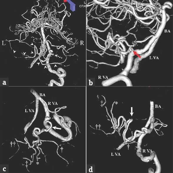
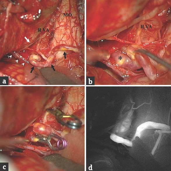
Although the anatomy of the posterior inferior cerebellar artery (PICA) is highly variable, a solitary PICA supplying both hemispheres of the cerebellum is rare. A 76-year-old woman presented with severe headache and subsequent loss of consciousness and was admitted to our hospital. Initial computed tomography showed subarachnoid hemorrhage. Three-dimensional digital subtraction angiography revealed a saccular aneurysm arising from the right vertebral artery (VA)-PICA bifurcation. The PICA branching from the right VA was enlarged, tortuous, and crossed the midline to supply both cerebellar hemispheres. This right PICA was interpreted as a bihemispheric PICA. Recognizing this variant preoperatively could help prevent complications of surgery. Careful follow-up studies are necessary in cases with bihemispheric PICA to monitor for the development of aneurysm at the junction between the bihemispheric PICA and the VA or the distal portion of the bihemispheric PICA.
Keywords: Aneurysm; bihemispheric posterior inferior cerebellar artery; subarachnoid hemorrhage; variant.
Conflict of interest statement
There are no conflicts of interest.
Figures
Similar articles
-
Successful endovascular treatment of a ruptured bihemispheric posterior inferior cerebellar artery aneurysm: illustrative case.J Neurosurg Case Lessons. 2021 Aug 16;2(7):CASE21367. doi: 10.3171/CASE21367. eCollection 2021 Aug 16. J Neurosurg Case Lessons. 2021. PMID: 35855413 Free PMC article.
-
Challenges in the Management of a Ruptured Bihemispheric Posterior Inferior Cerebellar Artery Aneurysm.World Neurosurg. 2019 Feb;122:317-321. doi: 10.1016/j.wneu.2018.11.051. Epub 2018 Nov 15. World Neurosurg. 2019. PMID: 30448579
-
The bihemispheric posterior inferior cerebellar artery.Neuroradiology. 2005 Nov;47(11):809-12. doi: 10.1007/s00234-005-1427-z. Epub 2005 Sep 14. Neuroradiology. 2005. PMID: 16160817
-
Delayed Establishment of Collateral Circulation from Posterior Meningeal Artery After Proximal Occlusion of Posterior Inferior Cerebellar Artery: Case Report and Literature Review.World Neurosurg. 2018 Jul;115:334-337. doi: 10.1016/j.wneu.2018.04.207. Epub 2018 May 9. World Neurosurg. 2018. PMID: 29751186 Review.
-
[A case of de novo aneurysm of the distal posterior inferior cerebellar artery with intraventricular hemorrhage].No Shinkei Geka. 1996 May;24(5):469-73. No Shinkei Geka. 1996. PMID: 8692375 Review. Japanese.
Cited by
-
A Prevalence Anatomic-Imaging Study of the Posterior Inferior Cerebellar Artery's Origin.Medicina (Kaunas). 2024 Aug 26;60(9):1397. doi: 10.3390/medicina60091397. Medicina (Kaunas). 2024. PMID: 39336438 Free PMC article.
-
Successful endovascular treatment of a ruptured bihemispheric posterior inferior cerebellar artery aneurysm: illustrative case.J Neurosurg Case Lessons. 2021 Aug 16;2(7):CASE21367. doi: 10.3171/CASE21367. eCollection 2021 Aug 16. J Neurosurg Case Lessons. 2021. PMID: 35855413 Free PMC article.
References
-
- Lasjaunias P, Vallee B, Person H, Ter Brugge K, Chiu M. The lateral spinal artery of the upper cervical spinal cord. Anatomy, normal variations, and angiographic aspects. J Neurosurg. 1985;63:235–41. - PubMed
-
- Carlson AP, Alaraj A, Dashti R, Aletich VA. The bihemispheric posterior inferior cerebral artery: Anatomic variations and clinical relevance in 11 cases. J Neurointerv Surg. 2013;5:601–4. - PubMed
-
- Cullen SP, Ozanne A, Alvarez H, Lasjaunias P. The bihemispheric posterior inferior cerebellar artery. Neuroradiology. 2005;47:809–12. - PubMed
-
- Reinacher P, Krings T, Burgel U, Hans FJ. Posterior inferior cerebellar artery (PICA) aneurysm arising from a bihemispheric PICA. Clin Neuroradiol. 2006;16:190–1.
Publication types
LinkOut - more resources
Full Text Sources
Other Literature Sources