

Intestinal epithelium, intraepithelial lymphocytes and the gut microbiota - Key players in the pathogenesis of celiac disease
- PMID: 29204051
- PMCID: PMC5698244
- DOI: 10.3748/wjg.v23.i42.7505
Intestinal epithelium, intraepithelial lymphocytes and the gut microbiota - Key players in the pathogenesis of celiac disease
Abstract
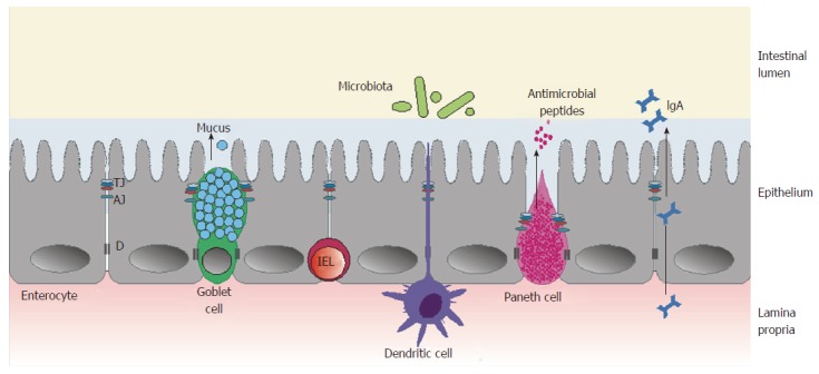
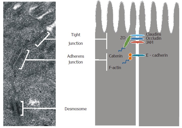
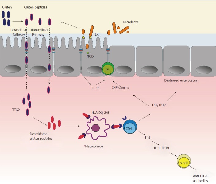
Celiac disease (CD) is a chronic immune-mediated disorder triggered by the ingestion of gluten in genetically predisposed individuals. Before activating the immune system, gluten peptides are transferred by the epithelial barrier to the mucosal lamina propria, where they are deamidated by intestinal tissue transglutaminase 2. As a result, they strongly bind to human leucocyte antigens (HLAs), especially HLA-DQ2 and HLA-DQ8, expressed on antigen-presenting cells. This induces an inflammatory response, which results in small bowel enteropathy. Although gluten is the main external trigger activating both innate and adaptive (specific) immunity, its presence in the intestinal lumen does not fully explain CD pathogenesis. It has been hypothesized that an early disruption of the gut barrier in genetically susceptible individuals, which would result in an increased intestinal permeability, could precede the onset of gluten-induced immune events. The intestinal barrier is a complex functional structure, whose functioning is dependent on intestinal microbiota homeostasis, epithelial layer integrity, and the gut-associated lymphoid tissue with its intraepithelial lymphocytes (IELs). The aim of this paper was to review the current literature and summarize the role of the gut microbiota, epithelial cells and their intercellular junctions, and IELs in CD development.
Keywords: Celiac disease; Epithelium; Intestinal barrier; Intestinal microbiota; Intraepithelial lymphocytes.
Conflict of interest statement
Conflict-of-interest statement: The authors declare no conflicts of interest.
Figures
Similar articles
-
How the Microbiota May Affect Celiac Disease and What We Can Do.Nutrients. 2024 Jun 14;16(12):1882. doi: 10.3390/nu16121882. Nutrients. 2024. PMID: 38931237 Free PMC article. Review.
-
Th17, intestinal microbiota and the abnormal immune response in the pathogenesis of celiac disease.Gastroenterol Hepatol Bed Bench. 2015 Spring;8(2):117-22. Gastroenterol Hepatol Bed Bench. 2015. PMID: 25926936 Free PMC article. Review.
-
Gliadin-dependent neuromuscular and epithelial secretory responses in gluten-sensitive HLA-DQ8 transgenic mice.Am J Physiol Gastrointest Liver Physiol. 2008 Jan;294(1):G217-25. doi: 10.1152/ajpgi.00225.2007. Epub 2007 Nov 15. Am J Physiol Gastrointest Liver Physiol. 2008. PMID: 18006603
-
Toxic, immunostimulatory and antagonist gluten peptides in celiac disease.Curr Med Chem. 2009;16(12):1489-98. doi: 10.2174/092986709787909613. Curr Med Chem. 2009. PMID: 19355902 Review.
-
The role of infectious mediators and gut microbiome in the pathogenesis of celiac disease.Arch Iran Med. 2015 Apr;18(4):244-9. Arch Iran Med. 2015. PMID: 25841946 Review.
Cited by
-
Simulated Gastric Digestion and In Vivo Intestinal Uptake of Orally Administered CuO Nanoparticles and TiO2 E171 in Male and Female Rat Pups.Nanomaterials (Basel). 2021 Jun 4;11(6):1487. doi: 10.3390/nano11061487. Nanomaterials (Basel). 2021. PMID: 34199726 Free PMC article.
-
Characterization of the "gut microbiota-immunity axis" and microbial lipid metabolites in atrophic and potential celiac disease.Front Microbiol. 2022 Sep 30;13:886008. doi: 10.3389/fmicb.2022.886008. eCollection 2022. Front Microbiol. 2022. PMID: 36246269 Free PMC article.
-
Lactobacillus rhamnosus GG Protects the Epithelial Barrier of Wistar Rats from the Pepsin-Trypsin-Digested Gliadin (PTG)-Induced Enteropathy.Nutrients. 2018 Nov 7;10(11):1698. doi: 10.3390/nu10111698. Nutrients. 2018. PMID: 30405050 Free PMC article.
-
The NLRP3 inflammasome in allergic diseases: mechanisms and therapeutic implications.Clin Exp Med. 2024 Sep 26;24(1):231. doi: 10.1007/s10238-024-01492-z. Clin Exp Med. 2024. PMID: 39325206 Free PMC article. Review.
-
Crosstalk between the oral microbiota, mucosal immunity, and the epithelial barrier regulates oral mucosal disease pathogenesis.Mucosal Immunol. 2021 Nov;14(6):1247-1258. doi: 10.1038/s41385-021-00413-7. Epub 2021 May 26. Mucosal Immunol. 2021. PMID: 34040155 Review.
References
Publication types
MeSH terms
LinkOut - more resources
Full Text Sources
Other Literature Sources
Medical
Research Materials
