Review
. 2017 Oct 31:11:224-282.
doi: 10.2174/1874285801711010224. eCollection 2017.

Paracoccidioidomycosis: Current Perspectives from Brazil

Affiliations
Review

Paracoccidioidomycosis: Current Perspectives from Brazil

Rinaldo Poncio Mendes et al. Open Microbiol J. .

Abstract

Background: This review article summarizes and updates the knowledge on paracoccidioidomycosis. P lutzii and the cryptic species of P. brasiliensis and their geographical distribution in Latin America, explaining the difficulties observed in the serological diagnosis.

Objectives: Emphasis has been placed on some genetic factors as predisposing condition for paracoccidioidomycosis. Veterinary aspects were focused, showing the wide distribution of infection among animals. The cell-mediated immunity was better characterized, incorporating the recent findings.

Methods: Serological methods for diagnosis were also compared for their parameters of accuracy, including the analysis of relapse.

Results: Clinical forms have been better classified in order to include the pictures less frequently observesiod.

Conclusion: Itraconazole and the trimethoprim-sulfamethoxazole combination was compared regarding efficacy, effectiveness and safety, demonstrating that azole should be the first choice in the treatment of paracoccidioidomycosis.

Keywords: Antifungal compounds; Endemic mycosis; Paracoccidioides brasiliensis; Paracoccidioides lutzii; Paracoccidioidmycosis; Systemic mycosis.


Figures

Fig. (1)
Fig. (1)
Paracoccidioides brasiliensis. A – a 10% KOH preparation of sputum showing multiple budding yeast forms, with birefringent walls; B – cell-block preparation of sputum (GMS, x400) showing the mickey-mouse and pilot will; C – histopathological examination showing granulomatous lesions with fungi within multinucleated giant cells (HE, x160); D – histopathological examination showing multiple budding yeast form (GMS x160). [B,C,D courtesy of the Department of Pathology – Faculdade de Medicina de Botucatu – UNESP].
Fig. (2)
Fig. (2)
Phylogeny and recombination in Paracoccidioides. Two methods were used to examine strain relationships originating from across South America (A): using 614,570 SNPs, including a phylogenetic network constructed with SplitsTree4 (B), and a Bayesian calibrated phylogeny constructed with BEAST (C); bootstrap values from maximum likelihood phylogeny constructed with RAxML were included for major subdivisions. Both methods show evidence of five distinct lineages in P. brasiliensis: S1 (blue), which is divided into two groups S1a (dark blue) and S1b (light blue), PS2 (green), PS3 (red), and the recently described PS4 (purple). Also, this phylogeny supports the divergence between P. brasiliensis and P. lutzii (Pl [orange]) as a different species. In addition, the phylogenetic network of P. brasiliensis suggests patterns of recombination (red branches). Note: from Muñoz JF, Farrer RA, Desardins CA, et al. Genome diversity, recombination, and virulence across the major lineages of Paracoccidioides. 2016. mSphere 1(5): e00213-16.
Fig. (3)
Fig. (3)
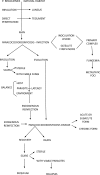
Natural history of paracoccidioidomycosis. Note: from Franco M, Mendes RP, Moscardi-Bacchi M, Rezkallah-Iwasso M, Montenegro MR. Paracoccidioidomycosis. Baillière’s Clin Trop Med Commun Dis. 1989; 4.1: 185 – 220.
Fig. (4)
Fig. (4)
Overview of the polarization of adaptive immune in PCM.(A) Activation of T helper (Th) subsets. Paracoccidioides antigen-primed dendritic cells (DCs) migrate to the lymph node where they present processed antigens to naïve Thcells that differentiate into one of Th lymphocyte subsets (Th1, Th2, Th9 and Th17) depending primarily on cytokines present in the extracellular environment. For Th1 polarization, IL-12 from DCs and macrophages, and IFN-γ from NK cells activate STAT1/STAT4 signaling to induce expression of the Th1-specific transcription factor, T-bet. For Th17 polarization, IL-6, IL-1β, TGF-β and IL-23 are required to induce expression of the Th17-specific transcription factor, RORβτ, through STAT3 signaling. For Th2 polarization, IL-4 from DCs activates STAT6 signaling to induce expression of the Th2-specific transcription factor, GATA-3. For Th9 polarization, IL-4 and TGF-β are required to induce expression of the Th9-specific transcription factor, PU.1. (B) Effector phases of T cell responses. Th1, Th17, Th2 and Th9 clones could be distinguished mainly by the cytokines produced by the cells. Th1 cells release high amounts of IFN-γ and TNF-α that classically activate macrophages (M1) resulting in fungal elimination. Th17 cells secrete IL-17 and IL-22 that recruit neutrophils and monocytes. Neutrophils cells act by generate reactive oxygen species (ROS), release of neutrophil extracellular traps (NETs) that result in fungal elimination. Monocytes have been studied in PCM by induce high levels of inflammatory cytokines, such as TNF-α and IL-1β, and growth factors, such as TGF-β and fibroblast growth factor (FGF). Th2 show several functions that depend of each secreted cytokine. IL-4 induces activation of B cells and subsequent production of immunoglobulins; IL-5 triggers recruitment of eosinophils; and IL-13 is involved in the deactivation of macrophages, termed “alternatively activated macrophages” (M2), that results in fungal growth and also in tissue repair. Th9 release IL-9 and IL-21 that act in synergy with Th2 to produce antibodies.
Fig. (5)
Fig. (5)
Proposed overview of fibrogenesis in PCM. The uncontrolled deposition of collagen fibers (types I and III) during a reparative and/or reactive process is recognized as fibrosis and the fibroblasts are the main cell involved in the production of collagens upon stimulation. During prolonged chronic inflammation, macrophages are classically activated (M1) by Th1 cells, pro-inflammatory cytokines and/orimmunocomplexes. These cells show intense production of cytotoxic metabolites to kill pathogens, release pro-inflammatory cytokines, induce tissue necrosis, and recruit phagocytes. Fresh monocytes from peripheral blood and monocyte-derived macrophages release inflammatory cytokines and growth factor that result inmacrophage activationsand fibroblast differentiation. Simultaneously, due to the constant tissue damage, others macrophages are alternatively activated (M2) to stimulatetissue repair. These cells promote the elimination of cellular debris, activation of fibroblast and down-regulate the activities of metalloproteinases, enzymes implicated in extracellular matrix/collagen degradation, promoting deposition of collagen fibers. On the other hand, M1-polarized macrophages up-regulate metalloproteinase activities, resulting in less collagen deposition. Besides the non-regulated, constant and prolonged reparative and reactive processes involved in fibrogenesis, do not rule out the action of molecules of Paracoccidioides in the activation of fibroblast.
Fig. (6)
Fig. (6)
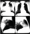
Chest radiograph of a patient with the chronic form of paracoccidioidomycosis, in antero-psterior and laterak view, showing bilateral and symmetrical alveolar lesions before (A and C) and after (B and D) treatment.
Fig. (7)
Fig. (7)
A - Paracoccidioidomycosis: infiltrated erythematous and well demarcated lesions on face; B - Paracoccidioidomycosis: compact granuloma with epithelioid and giant cells. A fungal cell is well observed in the cytoplasm of a giant cell (HE x40).
Fig. (8)
Fig. (8)
A - Paracoccidioidomycosis: ulcerated lesion with hemorrhagic dots and elevated borders on the face; B - Paracoccidioidomycosis: fungal cells stained in black, some in active multiplication. (Grocott-Gomori x 40); C - paracoccidioidomycosis: marked pseudoepitheliomatous hyperplasia with dense inflammatory infiltration in the dermis. Fungal cells can be observed. (HE x10).
Fig. (9)
Fig. (9)
Schedule of treatment and follow-up of patients with paracoccidioidomycosis. DID - serological evaluation performed by the double agar gel immunodiffusion test, as the inverse of the dilution. CMI – evaluation of the cell mediated immunity.
Fig. (10)
Fig. (10)
Patient with the acute/subacute form of paracoccidioidomycosis. A – lymph-node enlargement of the suppurative type before treatmnent; B – the same patient after treatment with itraconazole.

References

    1. Lutz A. Uma mycose pseudococcidica localisada na bocca e observada no Brazil. Contribuição ao conhecimento das hiphoblastomycoses americanas. Brazil-méd. 1908;22(121-124):141–144.
    1. Splendore A. Zymonematosi com localizzazione nella cavità della bocca, osservata in Brasile. Bull. Soc. Pathol. Exot. 1912;5:313–319.
    1. Almeida FP. Estudos comparativos de granuloma coccidióidico nos Estados Unidos e no Brasil: novo gênero para o parasito brasileiro. An Fac Med S Paulo. 1930;5:125–141.
    1. Jordan E.P. Standard nomenclature of diseases and standard nomenclature of operations. Chicago: American Medical Association; 1942.
    1. Paracoccidioidomycosis. Proceedings of the first pan american symposium, medellín (Colombia). PAHO Sci Publ. 1971;254

LinkOut - more resources