

Pediatric adrenal trauma: evaluation and follow-up with contrast-enhanced ultrasound (CEUS)
- PMID: 29204237
- PMCID: PMC5698183
- DOI: 10.1007/s40477-016-0220-7
Pediatric adrenal trauma: evaluation and follow-up with contrast-enhanced ultrasound (CEUS)
Abstract
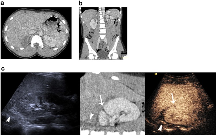
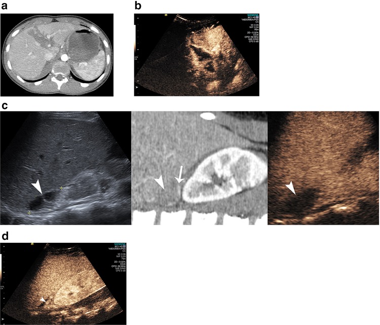
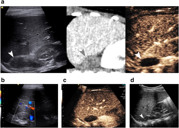
Adrenal trauma is rarely encountered in the pediatric population, often as a component of multi-organ injury and often presenting with hematoma formation. Contrast-enhanced computed tomography (CECT) is the established imaging modality both for initial evaluation and follow-up of patients with blunt abdominal trauma. In children, radiation exposure should be minimized and alternative imaging strategies are needed. Contrast-enhanced ultrasound (CEUS) has recently been successfully used for evaluation of patients with hepatic, renal and splenic injury. We present three cases of children with post-traumatic adrenal hematomas, which were initially diagnosed with CECT and subsequently followed up with CEUS, suggesting that CEUS may be considered for follow-up of adrenal injuries in children. CEUS improves the diagnostic capabilities by increasing tissue contrast and spatial resolution, readily differentiating viable from necrotic tissue in comparison to conventional ultrasound without the risk of iodine contrast agents and ionizing radiation of repeated CECT examinations.
Il trauma dei surreni accade raramente nella popolazione pediatrica, spesso è parte di un poli-trauma e spesso si presenta normalmente sotto forma di ematoma. La tomografia computerizzata con contrasto (TAC) è stabilita come modalità di diagnostica per immagini per la valutazione iniziale e il follow-up di pazienti con trauma addominale chiuso. Nei bambini e’ necessario ridurre al minimo l’esposizione alle radiazioni ionizzanti, pertanto sono necessarie strategie d’immagine alternative. L’ecografia con mezzo di contrasto (CEUS) e’ stata usata recentemente con successo nella valutazione di pazienti con lesioni traumatiche epatiche, renali e spleniche. Presentiamo tre casi di bambini con ematomi surrenali post-traumatici, inizialmente diagnosticati con tomografia computerizzata con contrasto (TAC) e successivamente esaminati al follow-up con ecografia con contrasto (CEUS), e suggeriamo che la CEUS può essere considerata per il follow-up delle lesioni traumatiche dei surreni nei bambini. La CEUS migliora le capacità diagnostiche aumentando contrasto tissutale e la risoluzione spaziale, differenziando prontamente il tessuto vitale da quello necrotico rispetto all’ecografia convenzionale, senza il rischio inerente ai mezzi di contrasto iodati e alle radiazioni ionizzanti legate a ripetuti esami con TAC con contrasto.
Keywords: Adrenal; Microbubbles; Pediatric; Trauma; Ultrasound.
Conflict of interest statement
Conflict of interest
Author AD has received lecture fees from Bracoo company. Author MES has received lecture fees from Bracco company. Author PSS has received lecture fees from Siemens, Hitachi, GE, Bracco companies.
Ethical approval
For this type of study formal consent is not required.
Human and animal studies
This article does not contain any studies with human participants or animals performed by any of the authors.
Informed consent
Informed consent was obtained from all individual participants included in the study.
Figures
References
-
- Claudon M, Dietrich CF, Choi BI, et al. Guidelines and good clinical practice recommendations for contrast enhanced ultrasound (CEUS) in the liver–update 2012: a WFUMB-EFSUMB initiative in cooperation with representatives of AFSUMB, AIUM, ASUM, FLAUS and ICUS. Ultraschall Med. 2013;34:11–29. - PubMed
Publication types
MeSH terms
Substances
LinkOut - more resources
Full Text Sources
Other Literature Sources
