

Wilkie's syndrome
- PMID: 29204239
- PMCID: PMC5698186
- DOI: 10.1007/s40477-017-0257-2
Wilkie's syndrome
Abstract
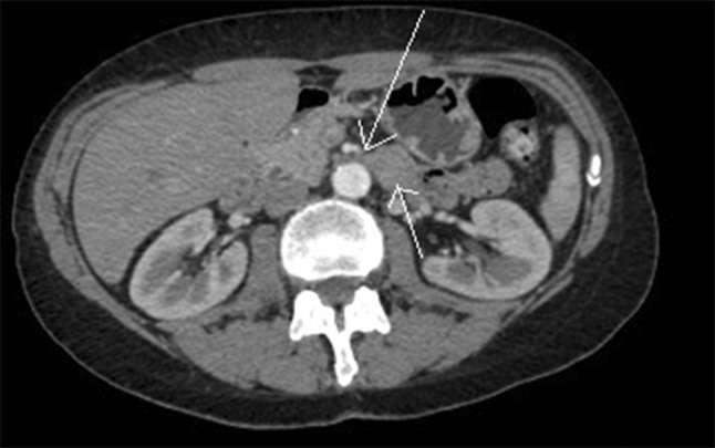
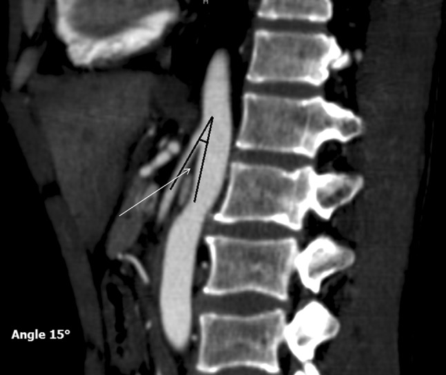
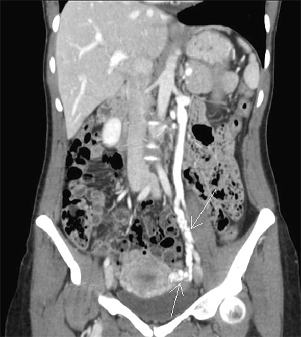
Superior mesenteric artery syndrome, also known as Wilkie's syndrome, is a rare vascular disease caused by the anomalous course of the superior mesenteric artery arising from the abdominal aorta with a smaller angle than the norm (<22°). The reduced angle compresses the structures situated between the aorta and the superior mesenteric artery, such as the duodenum and left renal vein; this can determine painful crises, intestinal subocclusions, and left varicocele. This syndrome can be congenital or acquired. The acquired type is more common and is generally caused by reduced perivascular fat surrounding the abdominal aorta and the superior mesenteric artery; this form is common among anorexic patients that have had a rapid weight loss. We present the case of a female patient who suffered from repeated postprandial vomiting and who lost 12 kg in 4 months. B-mode ultrasound imaging revealed evidence of a reduced angle between the aorta and the superior mesenteric artery, as found in Wilkie's syndrome. After diagnosis, the patient followed a high-calorie diet, and 2 months later an ultrasound scan proved the restoration of the aorto-mesenteric angle as a consequence of increased perivascular fat with regression of symptoms.
La sindrome dell’arteria mesenterica superiore o sindrome di Wilkie è una patologia vascolare rara dovuta all’anomalo decorso dell’arteria mesenterica superiore che nasce dall’aorta addominale con un angolo ridotto rispetto alla norma (inferiore a 22°). L’angolazione ridotta provoca la compressione delle strutture che passano tra l’aorta e l’arteria mesenterica superiore, il duodeno e la vena renale sinistra; questo può determinare crisi dolorose, sub-occlusioni intestinali e varicocele sinistro. Questa sindrome può essere congenita o acquisita. La forma acquisita, più frequente, è dovuta in genere alla riduzione del pannicolo adiposo peri-vascolare che circonda l’aorta addominale e l’arteria mesenterica superiore; questa forma è comune soprattutto nei pazienti anoressici che hanno subito una rapida perdita di peso. Presentiamo un caso di una paziente affetta da crisi ripetute di vomito post-prandiale che ha subito una perdita di 12 kg in 4 mesi. Durante l’esame ecografico B-Mode si evidenziava una riduzione dell’angolo tra aorta ed arteria mesenterica superiore, tipico della sindrome di Wilkie. Successivamente la paziente ha eseguito una dieta ipercalorica e dopo due mesi l’ecografia dimostrava il ripristino dell’angolo aorto-mesenterico come conseguenza dell’aumento del grasso peri-vascolare con regressione dei sintomi.
Keywords: Superior mesenteric artery; Ultrasound; Wilkie’s syndrome.
Conflict of interest statement
Conflict of interest
The authors declare that they have no conflict of interest.
Ethical approval
All procedures performed in studies involving human participants were in accordance with the ethical standards of the institutional and/or national research committee and with the 1964 Helsinki declaration and its later amendments or comparable ethical standards.
Informed consent
Informed consent was obtained from all individuals participating in the study.
Figures


References
-
- Wilkie DPD. Chronic duodenal ileus. Am J Med Sci. 1927;173:643. doi: 10.1097/00000441-192705000-00006. - DOI
Publication types
MeSH terms
LinkOut - more resources
Full Text Sources
Other Literature Sources
