

Acid suppressive therapy improved symptoms due to circumferential cervical inlet patch with proton pumps (H+/K+-ATPase)
- PMID: 29204429
- PMCID: PMC5700390
- DOI: 10.12998/wjcc.v5.i11.403
Acid suppressive therapy improved symptoms due to circumferential cervical inlet patch with proton pumps (H+/K+-ATPase)
Abstract
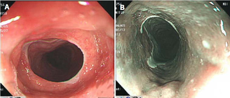
Cervical inlet patch (CIP), also referred to as esophageal heterotopic gastric mucosa, is regarded as the residue of columnar epithelium of the embryonic esophagus. Narrow band imaging increases the detection rate of CIP. Herein, we present a 55-year-old man with symptomatic circumferential inlet patch. He exhibited globus and dysphagia, and esophagogastroduodenoscopy found cir-cumferential CIP, where im-munohistochemistry revealed the existence of pro-ton pumps (H+, K+-ATPase). His throat symptoms were relieved by acid suppressive therapy with pump inhibitors. This case indicated that CIP should be considered as a differential diagnosis for the cause of globus symptoms in rare cases.
Keywords: Cervical inlet patch; Proton pump inhibitor.
Conflict of interest statement
Conflict-of-interest statement: No conflict interest to declare.
Figures

Similar articles
-
Intentional examination of esophagus by narrow-band imaging endoscopy increases detection rate of cervical inlet patch.Dis Esophagus. 2015 Oct;28(7):666-72. doi: 10.1111/dote.12252. Epub 2014 Jul 24. Dis Esophagus. 2015. PMID: 25059461
-
Esophageal Stricture: An Uncommon Complication of Cervical Inlet Patch.J Investig Med High Impact Case Rep. 2023 Jan-Dec;11:23247096231201024. doi: 10.1177/23247096231201024. J Investig Med High Impact Case Rep. 2023. PMID: 37840294 Free PMC article.
-
Prevalence and Clinical and Endoscopic Characteristics of Cervical Inlet Patch (Heterotopic Gastric Mucosa): A Systematic Review and Meta-Analysis.J Clin Gastroenterol. 2022 Mar 1;56(3):e250-e262. doi: 10.1097/MCG.0000000000001516. J Clin Gastroenterol. 2022. PMID: 33780217
-
Symptoms of Chronic Dysphagia Secondary to Multiple Cervical Inlet Patches and Esophageal Stricture.Cureus. 2023 Jan 6;15(1):e33459. doi: 10.7759/cureus.33459. eCollection 2023 Jan. Cureus. 2023. PMID: 36751259 Free PMC article.
-
Cervical inlet patch: new insights into diagnosis and endoscopic therapy.Frontline Gastroenterol. 2018 Jul;9(3):214-220. doi: 10.1136/flgastro-2017-100855. Epub 2017 Nov 9. Frontline Gastroenterol. 2018. PMID: 30046427 Free PMC article. Review.
Cited by
-
Heterotopic Gastric Mucosa in Middle Esophagus Complicated with Esophageal Ulcers.Intern Med. 2022 Sep 15;61(18):2735-2740. doi: 10.2169/internalmedicine.8705-21. Epub 2022 Feb 26. Intern Med. 2022. PMID: 35228416 Free PMC article.
-
Issues and controversies in esophageal inlet patch.World J Gastroenterol. 2019 Aug 14;25(30):4061-4073. doi: 10.3748/wjg.v25.i30.4061. World J Gastroenterol. 2019. PMID: 31435164 Free PMC article. Review.
-
A systematic review of fully circumferential inlet patches (heterotopic gastric mucosa): More complicated than regular inlet patches.Indian J Gastroenterol. 2025 Aug;44(4):443-456. doi: 10.1007/s12664-025-01738-y. Epub 2025 Mar 31. Indian J Gastroenterol. 2025. PMID: 40163316 Free PMC article.
-
Esophageal Polyp Lesion in a Patient with Globus Pharyngeus Compliant.Middle East J Dig Dis. 2020 Jan;12(1):52-54. doi: 10.15171/mejdd.2020.165. Middle East J Dig Dis. 2020. PMID: 32082523 Free PMC article. No abstract available.
References
-
- von Rahden BH, Stein HJ, Becker K, Liebermann-Meffert D, Siewert JR. Heterotopic gastric mucosa of the esophagus: literature-review and proposal of a clinicopathologic classification. Am J Gastroenterol. 2004;99:543–551. - PubMed
-
- Hori K, Kim Y, Sakurai J, Watari J, Tomita T, Oshima T, Kondo C, Matsumoto T, Miwa H. Non-erosive reflux disease rather than cervical inlet patch involves globus. J Gastroenterol. 2010;45:1138–1145. - PubMed
-
- Galan AR, Katzka DA, Castell DO. Acid secretion from an esophageal inlet patch demonstrated by ambulatory pH monitoring. Gastroenterology. 1998;115:1574–1576. - PubMed
Publication types
LinkOut - more resources
Full Text Sources
Other Literature Sources
