

A 3D benign paroxysmal positional vertigo model for study of otolith disease
- PMID: 29204541
- PMCID: PMC5698514
- DOI: 10.1016/j.wjorl.2016.02.002
A 3D benign paroxysmal positional vertigo model for study of otolith disease
Abstract
Objective: To develop a three-dimensional study tool of the membranous labyrinth in order to study the pathophysiology, diagnostic workup and treatment of benign paroxysmal positional vertigo (BPPV). BPPV is the most common cause of peripheral vertigo. Its diagnosis and treatment depend on an understanding of the anatomy of the vestibular labyrinth and its position relative to the head. To date, many illustrations have been made to explain principals of diagnosis and treatment of BPPV, but few have been based on anatomical studies of the membranous labyrinth.
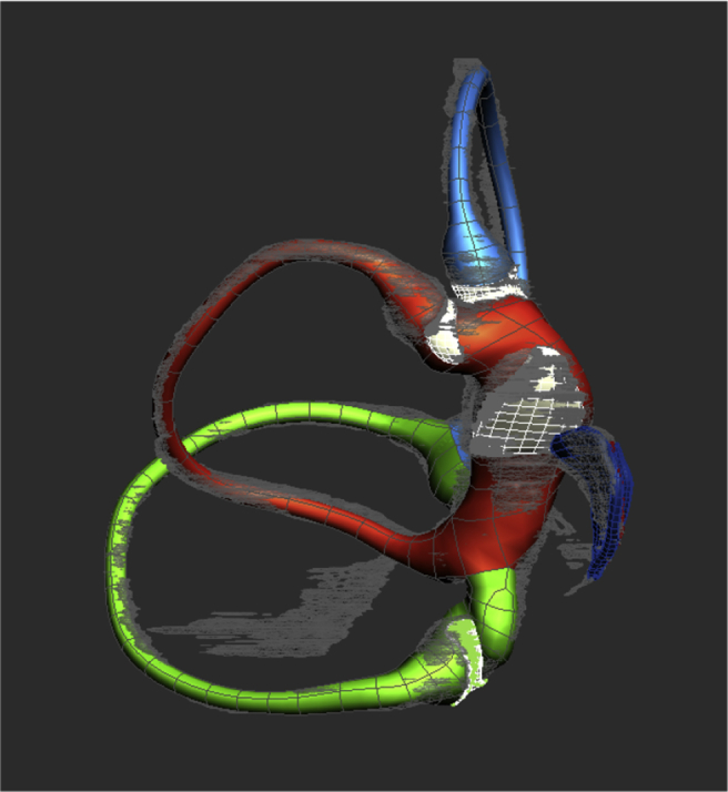
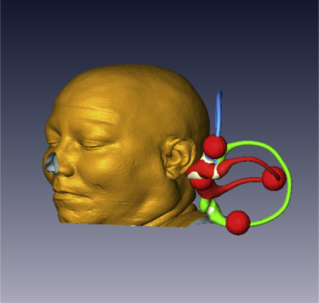
Methods: A cadaveric human membranous labyrinth was axially sectioned at 20 μm resolution, stained and segmented to create a high-resolution digital model. The model was cloned to create an enantiomeric pair of labyrinths. These were associated a 3D model of a human skull, segmented from MRI data, and were oriented according to established anatomic norms. Canal markers representing otoliths were created to mark canalith position during movement of the model within the 3D environment.
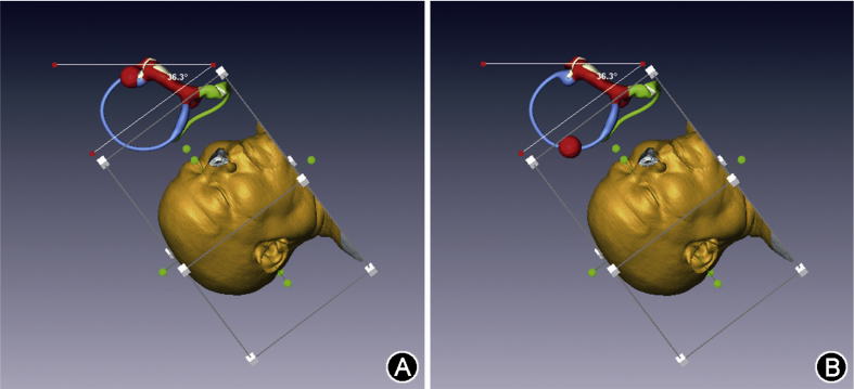
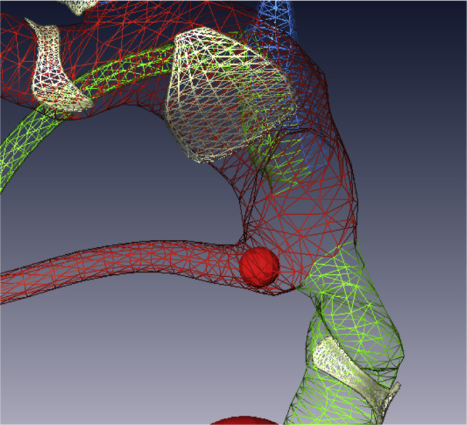
Results: The model allows visualization of true membranous labyrinth anatomy in both ears simultaneously. The dependent portion of each semicircular duct and of the utricle can easily be visualized in any head position. Moveable markers can mark the expected progress of otolith debris with changes in head position and images can be captured to document simulations. The model can be used to simulate pathology as well as diagnostic maneuvers and treatment procedures used for BPPV. The model has great potential as a teaching tool.
Conclusion: A simple model based on human anatomy has been created to allow careful study of BPPV pathophysiology and treatment. Going forward, this tool could offer insights that may lead to more accurate diagnosis and treatment of BPPV.
Keywords: 3D; Anatomy; Benign paroxysmal positional vertigo; Histology; Model; Modeling.
Figures


References
-
- Schuknecht H.F. Cupulolithiasis Arch Otolaryngol. 1969;90:765–778. - PubMed
-
- Epley J.M. The canalith repositioning procedure: for treatment of benign paroxysmal positional vertigo. Otolaryngol Head Neck Surg. 1992;107:399–404. - PubMed
-
- Hashimoto S., Naganuma H., Tokumasu K., Itoh A., Okamoto M. Three-dimensional reconstruction of the human semicircular canals and measurement of each membranous canal plane defined by Reid's stereotactic coordinates. Ann Otol Rhinol Laryngol. 2005;114:934–938. - PubMed
-
- Siebenmann F. Bergmann; Wiesbaden: 1890. Die Korrosions-Anatomie des knˆchernen Labyrinthes des menschlichen ohres.
LinkOut - more resources
Full Text Sources
Other Literature Sources
