

Advancing theranostics with tumor-targeting peptides for precision otolaryngology
- PMID: 29204554
- PMCID: PMC5698525
- DOI: 10.1016/j.wjorl.2016.05.006
Advancing theranostics with tumor-targeting peptides for precision otolaryngology
Abstract
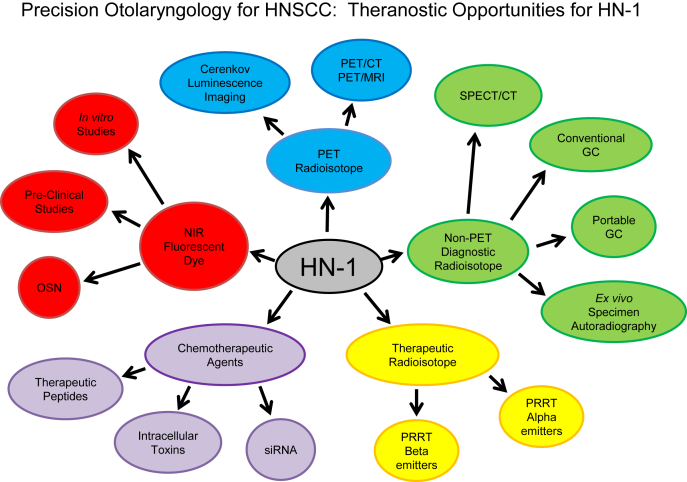
Worldwide, about 600,000 head and neck squamous cell carcinoma (HNSCC) are detected annually, many of which involve high risk human papilloma virus (HPV). Surgery is the primary and desired first treatment option. Following surgery, the existence of cancer cells at the surgical margin is strongly associated with eventual recurrence of cancer and a poor outcome. Despite improved surgical methods (robotics, microsurgery, endoscopic/laparoscopic, and external imaging), surgeons rely only on their vision and touch to locate tumors during surgery. Diagnostic imaging systems like computed tomography (CT), magnetic resonance imaging (MRI), single-photon emission computed tomography (SPECT) and positron-emission tomography (PET) are too large, slow and costly to use efficiently during most surgeries and, ultrasound imaging, while fast and portable, is not cancer specific. This purpose of this article is to review the fundamental technologies that will radically advance Precision Otolaryngology practices to the benefit of patients with HNSCC. In particular, this article will address the potential for tumor-targeting peptides to enable more precise diagnostic imaging while simultaneously advancing new therapeutic paradigms for next generation image-guided surgery, tumor-specific chemotherapeutic delivery and tumor-selective targeted radiotherapy (i.e., theranostic).
Keywords: Diagnostic imaging; Optical surgical navigation; Peptide; Squamous cell carcinoma; Theranostic.
Figures
Similar articles
-
Diagnostic performance of 18 fluorodesoxyglucose positron emission/computed tomography and magnetic resonance imaging in detecting T1-T2 head and neck squamous cell carcinoma.Laryngoscope. 2018 Feb;128(2):378-385. doi: 10.1002/lary.26729. Epub 2017 Jun 10. Laryngoscope. 2018. PMID: 28600822
-
More advantages in detecting bone and soft tissue metastases from prostate cancer using 18F-PSMA PET/CT.Hell J Nucl Med. 2019 Jan-Apr;22(1):6-9. doi: 10.1967/s002449910952. Epub 2019 Mar 7. Hell J Nucl Med. 2019. PMID: 30843003
-
Positron emission tomography-computed tomography versus positron emission tomography-magnetic resonance imaging for diagnosis of oral squamous cell carcinoma: A pilot study.J Craniomaxillofac Surg. 2015 Dec;43(10):2129-35. doi: 10.1016/j.jcms.2015.08.030. Epub 2015 Sep 11. J Craniomaxillofac Surg. 2015. PMID: 26498514
-
Current and Future Approaches for Effective Cancer Imaging and Treatment.Cancer Biother Radiopharm. 2018 Mar;33(2):39-51. doi: 10.1089/cbr.2017.2378. Cancer Biother Radiopharm. 2018. PMID: 29634415 Review.
-
[Current oncologic concepts and emerging techniques for imaging of head and neck squamous cell cancer].Laryngorhinootologie. 2012 Mar;91 Suppl 1:S27-47. doi: 10.1055/s-0031-1297259. Epub 2012 Mar 28. Laryngorhinootologie. 2012. PMID: 22456918 Review. German.
Cited by
-
Novel Peptide NIRF Optical Surgical Navigation Agents for HNSCC.Molecules. 2019 Aug 23;24(17):3070. doi: 10.3390/molecules24173070. Molecules. 2019. PMID: 31450798 Free PMC article.
-
Graphene oxide loaded with tumor-targeted peptide and anti-cancer drugs for cancer target therapy.Sci Rep. 2021 Jan 18;11(1):1725. doi: 10.1038/s41598-021-81218-3. Sci Rep. 2021. PMID: 33462277 Free PMC article.
-
Biological Evaluation of a Fluorescent-Imaging Agent for Medullary Thyroid Cancer in an Orthotopic Model.J Clin Endocrinol Metab. 2017 Sep 1;102(9):3268-3277. doi: 10.1210/jc.2017-00573. J Clin Endocrinol Metab. 2017. PMID: 28591772 Free PMC article.
-
Tongue squamous cell carcinoma-targeting Au-HN-1 nanosystem for CT imaging and photothermal therapy.Int J Oral Sci. 2025 Jan 14;17(1):9. doi: 10.1038/s41368-024-00343-7. Int J Oral Sci. 2025. PMID: 39805836 Free PMC article.
-
Tumor specifically internalizing peptide 'HN-1': Targeting the putative receptor retinoblastoma-regulated discoidin domain receptor 1 involved in metastasis.World J Clin Oncol. 2022 May 24;13(5):323-338. doi: 10.5306/wjco.v13.i5.323. World J Clin Oncol. 2022. PMID: 35662982 Free PMC article.
References
-
- Kamangar F., Dores G.M., Anderson W.F. Patterns of cancer incidence, mortality, and prevalence across five continents: defining priorities to reduce cancer disparities in different geographic regions of the world. J Clin Oncol. 2006;24:2137–2150. - PubMed
-
- Ramqvist T., Dalianis T. An epidemic of oropharyngeal squamous cell carcinoma (OSCC) due to human papillomavirus (HPV) infection and aspects of treatment and prevention. Anticancer Res. 2011;31:1515–1519. - PubMed
-
- Leemans C.R., Tiwari R., Nauta J.J., van der Waal I., Snow G.B. Recurrence at the primary site in head and neck cancer and the significance of neck lymph node metastases as a prognostic factor. Cancer. 1994;73:187–190. - PubMed
-
- Liao C.T., Chang J.T., Wang H.M. Analysis of risk factors of predictive local tumor control in oral cavity cancer. Ann Surg Oncol. 2008;15:915–922. - PubMed
-
- Leemans C.R., Tiwari R., Nauta J.J., van der Waal I., Snow G.B. Regional lymph node involvement and its significance in the development of distant metastases in head and neck carcinoma. Cancer. 1993;71:452–456. - PubMed
Publication types
Grants and funding
LinkOut - more resources
Full Text Sources
Other Literature Sources
