

Volumetric response of progressing post-SRS lesions treated with laser interstitial thermal therapy
- PMID: 29204838
- PMCID: PMC5823725
- DOI: 10.1007/s11060-017-2694-3
Volumetric response of progressing post-SRS lesions treated with laser interstitial thermal therapy
Erratum in
-
Correction to: Volumetric response of progressing post-SRS lesions treated with laser interstitial thermal therapy.J Neurooncol. 2019 Jan;141(2):475. doi: 10.1007/s11060-019-03093-3. J Neurooncol. 2019. PMID: 30635762
Abstract
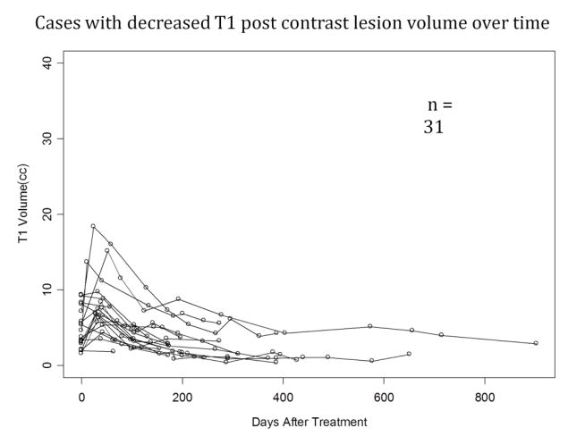
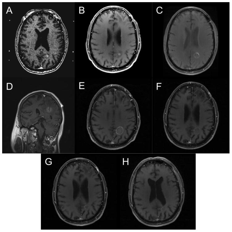
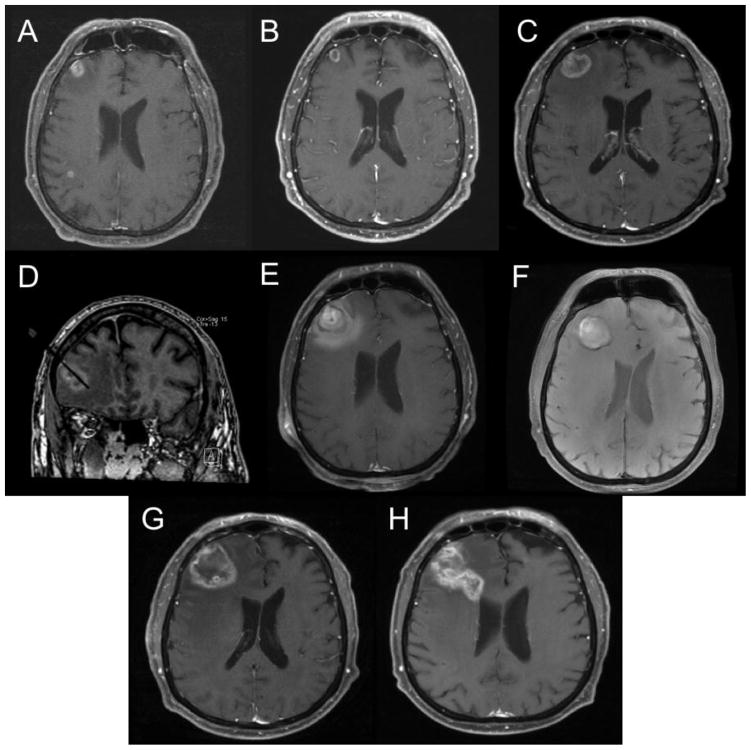
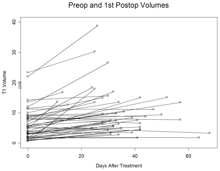
We analyzed volumetric response of metastatic brain tumors that progressed despite treatment with stereotactic radiosurgery (SRS) after treatment with laser interstitial thermal therapy (LITT). We retrospectively reviewed consecutive patients treated from 1/2012 to 10/2015 with LITT for metastatic brain tumors demonstrating progression after SRS. Volumes were quantified using MRI with contrast-enhanced T1-weighted (T1W) and fluid-attenuated inversion recovery (FLAIR). Fifty lesions from 36 patients were studied. Lesions were assessed prior to LITT, immediately after LITT, 0-90 days after LITT, 90-180 days after LITT, 180-270 days after LITT, and 270-360 days after LITT. The median T1W volume was 5.05 cc (range 0.54-23.31 cc) before LITT treatment (n = 50), 7.70 cc (range 1.72-38.76 cc) 0-90 days after LITT (n = 47), and 3.68 cc (range 1.282-48.31 cc) 180-270 days after LITT (n = 21). The median FLAIR volume was 43.36 cc (range 3.09-233.01 cc) before LITT treatment (n = 50), 37.13 cc (range 3.48-244.23 cc) 0-90 days after LITT (n = 43), 31.68 cc (range 1.6-248.75 cc) 180-270 days after LITT (n = 18). The 6-month FLAIR volume showed a statistically significant reduction compared to pretreatment (p = 0.04). After selecting for cases where patients had two or more post-operative MRIs, we found that 24 lesions (63%) demonstrated an overall downward trend and 14 lesions (37%) demonstrated an upward trend. The median pre-treatment T1W volume for the patients whose lesions demonstrated volumetric reduction after LITT was 3.54 cc (range 0.539-10.06 cc) and for those who did not demonstrate volumetric reduction after LITT it was 8.81 cc (range 0.926-23.313 cc). The pre-treatment tumor volume plays a significant role in determining response to LITT with smaller tumor volumes responding better to LITT than tumors with larger volumes.
Keywords: Brain metastasis; Laser interstitial thermal therapy; Metastatic; Post-SRS; Stereotactic radiosurgery.
Figures

References
-
- Carpentier A, McNichols RJ, Stafford RJ, Guichard JP, Reizine D, Delaloge S, Vicaut E, Payen D, Gowda A, George B. Laser thermal therapy: real-time MRI-guided and computer-controlled procedures for metastatic brain tumors. Lasers in surgery and medicine. 2011;43:943–950. doi: 10.1002/lsm.21138. - DOI - PubMed
-
- Carpentier A, McNichols RJ, Stafford RJ, Itzcovitz J, Guichard JP, Reizine D, Delaloge S, Vicaut E, Payen D, Gowda A, George B. Real-time magnetic resonance-guided laser thermal therapy for focal metastatic brain tumors. Neurosurgery. 2008;63:ONS21–28. doi: 10.1227/01.neu.0000335007.07381.df. discussion ONS28-29. - DOI - PubMed
-
- Mohammadi AM, Hawasli AH, Rodriguez A, Schroeder JL, Laxton AW, Elson P, Tatter SB, Barnett GH, Leuthardt EC. The role of laser interstitial thermal therapy in enhancing progression-free survival of difficult-to-access high-grade gliomas: a multicenter study. Cancer medicine. 2014;3:971–979. doi: 10.1002/cam4.266. - DOI - PMC - PubMed
MeSH terms
Grants and funding
LinkOut - more resources
Full Text Sources
Other Literature Sources
Medical
