Review
. 2018 Apr;10(2):481-492.
doi: 10.1007/s12551-017-0362-7. Epub 2017 Dec 4.

How multi-scale structural biology elucidated context-dependent variability in ectodomain conformation along with the ligand capture and release cycle for LDLR family members

Affiliations
Review

How multi-scale structural biology elucidated context-dependent variability in ectodomain conformation along with the ligand capture and release cycle for LDLR family members

Terukazu Nogi. Biophys Rev. 2018 Apr.

Abstract

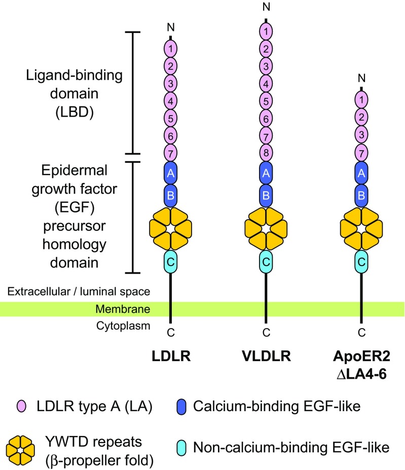
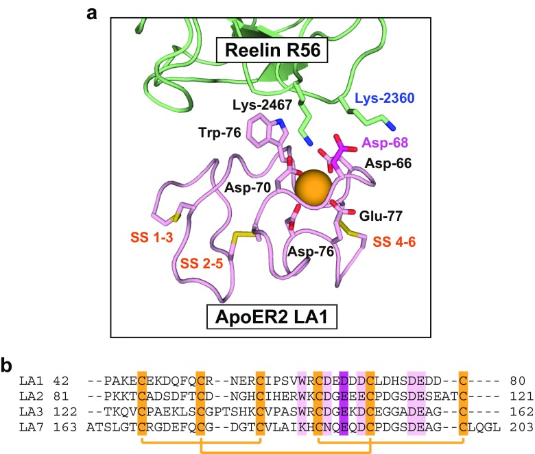
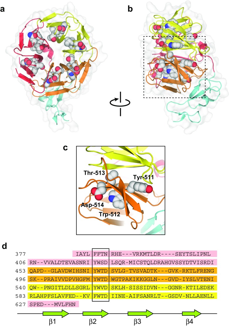
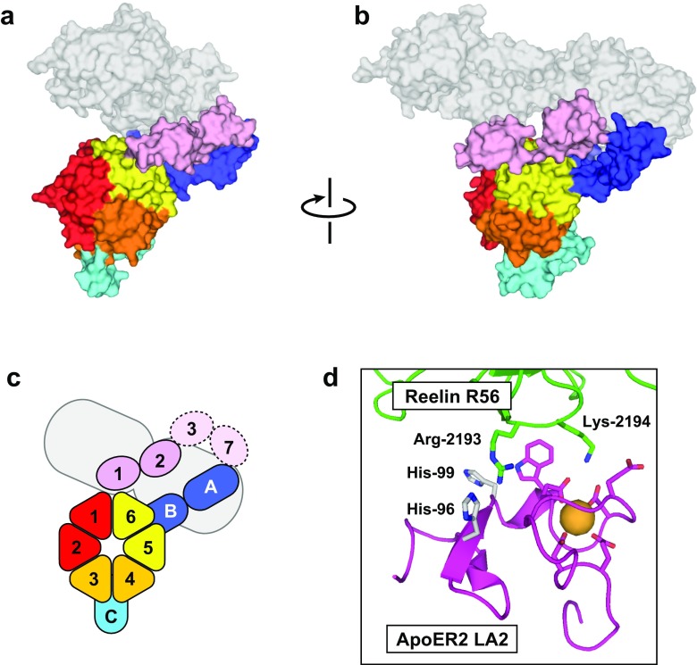
The low-density lipoprotein receptor (LDLR) and its homologs capture and internalize lipoproteins into the cell. Due to the fact that LDLR family members possess a modular ectodomain that undergoes dynamic conformational changes, multi-scale structural analysis has been performed so as to understand the ligand capture and release mechanism. For example, crystallographic analyses have provided models for both the entire ectodomain and high-resolution structures of individual modules. In addition, nuclear magnetic resonance spectroscopic analyses have shown the rigidity and flexibility of inter-module linkers to restrict the mobility of ectodomain. Accumulated structural data suggest that the ectodomains of LDLR family members are flexible at the cell surface and switch between two metastable conformations, that is, the extended and contracted conformations. Recent structural analysis of ApoER2, a close homolog of LDLR, raised the possibility that the receptor binds with the ligand in the contracted conformation. After transport to an endosome by endocytosis, the receptor undergoes a conformational change to the closed conformation for completion of ligand release. In contrast, LDLR has been reported to adopt the extended conformation when it binds with a inhibitory regulator that recruits LDLR toward the degradation pathway. These findings support a mechanism of different ectodomain conformations for binding the ligand versus binding the regulatory protein. In this review, I provide an overview of studies that analyze the structural and biophysical properties of the ectodomains of LDLR family members and discuss a hypothetical model for ligand uptake and receptor recycling that integrates the known ectodomain conformational variability.

Keywords: Conformational change; Endocytosis; Low-density lipoprotein receptor; Molecular recognition; X-ray crystallography.


Conflict of interest statement

Conflict of interest

Terukazu Nogi declares that he has no conflicts of interest.

Ethical approval

This article does not contain any studies with human participants or animals performed by the author.

Figures

Fig. 1
Fig. 1
Domain organization of human low-density lipoprotein receptor (LDLR) and its close homologs. The N-terminal domain of LDLR is termed as LBD while the C-terminal domain shows a homology to the EGF precursor. The number of LA modules differ between homologs, and the major splicing variant of human apolipoprotein E (ApoE)R2 contains four LA modules, lacking LA 4–6. The YWTD repeats assume a six-bladed β-propeller fold. The first and second EGF-like modules (EGF-A and -B) are classified as the calcium-binding type, and the third EGF-like module (EGF-C) is non-calcium-binding type. VLDLR Very-low-density lipoprotein receptor
Fig. 2
Fig. 2
Structure of the LA module in complex with a ligand. The crystal structure of the ApoE receptor 2 (ApoER2) N-terminal first LA (LA1) in complex with the reelin R56 fragment is shown as a representative example. a Close-up view of the crystal structure of ApoER2 LA1 in complex with the reelin R56 fragment. In ApoER2 LA1, the conserved residues constituting the ligand-binding pocket and disulfide bonds are shown as a stick model. The orange sphere represents the calcium ion coordinated by the conserved acidic residues. The two basic residues recognized by LA1 in a double-Lys/Arg mode are also shown in the stick model. b Sequence alignment of LA modules in human ApoER2 ΔLA4–6. The disulfide pairs are connected with orange lines. The conserved acidic and aromatic residues are indicated in pink. The acidic residues that do not coordinate the calcium ion but contribute to the electrostatic interactions are highlighted in dark magenta
Fig. 3
Fig. 3
Structure of the EGF-like modules and their interaction mode with proprotein convertase subtilisin/kexin type 9 (PCSK9). a The nuclear magnetic resonance structure of the LDLR–EGF-AB unit. EGF-A and -B, both calcium-binding EGF modules, are shown in light and dark colors, respectively. The residues contributing to the calcium-binding consensus and to the disulfide bonds are highlighted in stick model. Each EGF-like module contains a calcium ion, as shown in sphere model. EGF-A lacks the first consensus Asp/Asn residue corresponding to Asp-333 of EGF-B. b Close-up view of the PCSK9–EGF-A interface. PCSK9 docks to the surface close to the calcium-binding site of EGF-A. The side-chains of the residues involved in the inter-molecular interaction and the main-chains of the residues surrounding the calcium ion are shown in stick model. Since the structure was solved at acidic pH, the additional hydrogen bond was formed between His-306 of EGF-A and Asp-374 of PCSK9
Fig. 4
Fig. 4
Structure of the YWTD repeat and EGF-C, a non-calcium-binding EGF module. a Full view from the top-face of the β-propeller. b Full view from the side of the β-propeller. The structure is shown in ribbon model with a transparent surface. The residues in the YWTD consensus sequences are highlighted in sphere model. The six blades of the β-propeller are shown in the three different colors, and the EGF-C modules are shown in cyan. c Close-up view of the region around the fourth YWTD consensus. d Sequence alignment of the six blades of the β-propeller. The YWTD sequences form the second β-strand (β2), as highlighted with the box. The C-terminal region is incorporated into the blade containing the first YWTD sequence as the first β-strand (β1)
Fig. 5
Fig. 5
Structure of the entire ectodomain of LDLR. a Full view of the ligand-unbound LDLR ectodomain at acidic pH. b Close-up view of the intramolecular binding interface. The structure is shown in ribbon model with the calcium ions in sphere model. The residues involved in the intramolecular contacts are shown in stick model. Both LA4 and LA5 interact with the top-face of the β-propeller in the canonical ligand-binding mechanism. The protonated histidine residues, His-190, 560, and 582, are thought to stabilize the interactions. c Surface model of the ligand-unbound LDLR ectodomain at acidic pH. d Surface model of the PCSK9-bound LDLR ectodomain at neutral pH. PCSK9 is shown with transparent surface. e, f Diagram of the domain arrangement of ligand-unbound LDLR (e) and PCSK9-bound LDLR (f). The ligand-bound LDLR ectodomain adopts the contracted-closed conformation, in which the EGF-AB unit is located beside Blade 5–6, and draws the LA modules close to the top-face of β-propeller. In contrast, PCSK9-bound LDLR adopts the extended-open conformation, in which the EGF-AB unit is located beside Blade 1–2 and keeps the LA modules away from the β-propeller. The LA1–6 modules are outlined with a dotted line since they were not assigned in the crystal structure
Fig. 6
Fig. 6
Structure of the ApoER2 ectodomain in complex with the fragment containing the fifth and sixth repeats of reelin (R56). a Surface model viewed from the top-face of the β-propeller. b Side-view of the surface model. The reelin R56 fragment is shown as a transparent surface. c Diagram of the domain arrangement. Similar to ligand-unbound LDLR at acidic pH, the EGF-AB unit is located at the Blade 5–6 side (see text for description of “blade”). This conformation was defined as the contracted-open conformation as the LA modules are located close to the β-propeller, but they are still capable of binding with the ligand. d Close-up view of the binding interface between ApoER2 LA2 and reelin R56. The structure is shown in ribbon model, and the residues involved in the binding interfaces and calcium coordination are shown in stick model. The two histidine residues, His-96 and -99, on the binding surface of LA are presumed to contribute to the pH dependency of the binding interactions
Fig. 7
Fig. 7
Classification of domain arrangement in the EGF–YWTD–EGF unit from the LDLR family. a Structures with the incoming EGF at the Blade 5–6 side. b Structures with the incoming EGF beside Blades 1–2. The structures are shown in ribbon model with a transparent surface. The structures are viewed from the top-face of β-propeller, as shown in Figs. 5 and 6
Fig. 8
Fig. 8
Hypothetical model of ligand uptake and receptor recycling in the LDLR family members based on multi-scale structural biology. Presumably, the ectodomain of the receptor is flexible and can adopt both extended and contracted conformations at the cell surface. Structural analysis of ApoER2 raises the possibility that the receptor binds with the ligand in the contracted-open conformation, which seems to be primed for ligand release and adoption of the contracted-closed conformation when the receptor is recruited to the acidic endosomal compartment. In contrast, PCSK9 preferably binds with the receptor in the extended-open conformation. The interaction between the receptor and PCSK9 is stabilized in the endosome, and the receptor is subsequently transported to lysosome for degradation

Similar articles

Cited by

References

    1. Abifadel M, Varret M, Rabès JP, et al. Mutations in PCSK9 cause autosomal dominant hypercholesterolemia. Nat Genet. 2003;34:154–156. doi: 10.1038/ng1161. - DOI - PubMed
    1. Beglova N, Jeon H, Fisher C, Blacklow SC. Cooperation between fixed and low pH-inducible interfaces controls lipoprotein release by the LDL receptor. Mol Cell. 2004;16:281–292. doi: 10.1016/j.molcel.2004.09.038. - DOI - PubMed
    1. Bottomley MJ, Cirillo A, Orsatti L, et al. Structural and biochemical characterization of the wild type PCSK9-EGF(AB) complex and natural familial hypercholesterolemia mutants. J Biol Chem. 2009;284:1313–1323. doi: 10.1074/jbc.M808363200. - DOI - PubMed
    1. Brown MS, Goldstein JL. Expression of the familial hypercholesterolemia gene in heterozygotes: mechanism for a dominant disorder in man. Science. 1974;185:61–63. doi: 10.1126/science.185.4145.61. - DOI - PubMed
    1. Brown MS, Goldstein JL. A receptor-mediated pathway for cholesterol homeostasis. Science. 1986;232:34–47. doi: 10.1126/science.3513311. - DOI - PubMed