

Gliadins from wheat grain: an overview, from primary structure to nanostructures of aggregates
- PMID: 29204878
- PMCID: PMC5899726
- DOI: 10.1007/s12551-017-0367-2
Gliadins from wheat grain: an overview, from primary structure to nanostructures of aggregates
Abstract
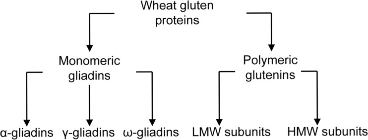
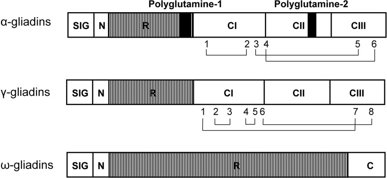
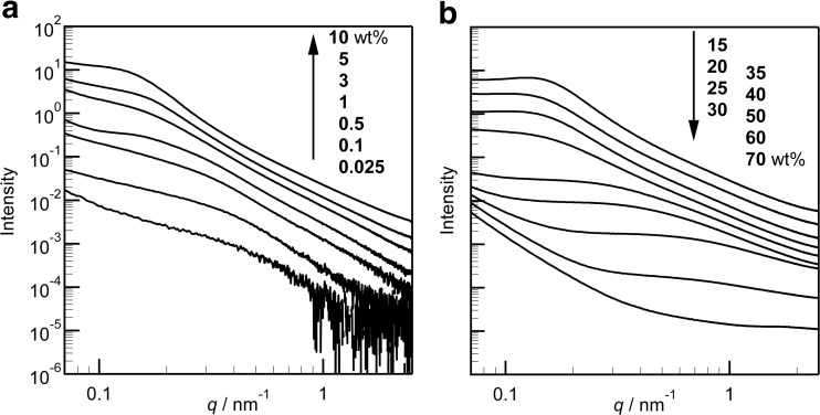
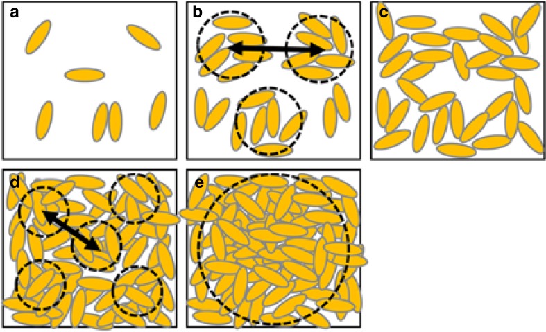
Gliadins are well-known wheat grain proteins, particularly important in food science. They were studied as early as the 1700s. Despite their long history, it has been difficult to identify their higher-order structure as they aggregate in aqueous solution. Consequently, most studies have been performed by extracting the proteins in 70% ethanol or dilute acidic solutions. The carboxy-terminal half of α- and γ-gliadins have α-helix-rich secondary structures stabilized with intramolecular disulfide bonds, which are present in either aqueous ethanol or pure water. The amino-terminal-repeat region of α- and γ-gliadins has poly-L-proline II and β-reverse-turn structures. ω-Gliadins also have poly-L-proline II and β-reverse-turn structures, but no α-helix structure. The size and shape of gliadin molecules have been determined by assessing a variety of parameters: their sedimentation velocity in the analytical ultracentrifuge, intrinsic viscosity, small-angle X-ray scattering profile, and images of the proteins from scanning probe microscopes such as a tunneling electron microscope and atomic force microscope. Models for gliadins are either rods or prolate ellipsoids whether in aqueous ethanol, dilute acid, or pure water. Recently, gliadins have been shown to be soluble in pure water, and a novel extraction method into pure water has been established. This has made it possible to analyze gliadins in pure water at neutral pH, and permitted the characterization of hydrated gliadins. They formed hierarchical nanoscale structures with internal density fluctuations at high protein concentrations.
Keywords: Gliadin; Nanostructure; Protein aggregate; SAXS; Wheat protein.
Conflict of interest statement
Conflict of interest
Reiko Urade declares that she has no conflicts of interest. Nobuhiro Sato declares that he has no conflicts of interest. Masaaki Sugiyama declares that he has no conflicts of interest.
Ethical approval
This article does not contain any studies with human participants or animals performed by any of the authors.
Figures
References
-
- Altenbach SB, Kothari KM. Omega gliadin genes expressed in Triticum aestivum cv. Butte 86: effects of post-anthesis fertilizer on transcript accumulation during grain development. J Cereal Sci. 2007;46:169–177. doi: 10.1016/j.jcs.2007.02.001. - DOI
-
- Altschuler Y, Galili G. Role of conserved cysteines of a wheat gliadin in its transport and assembly into protein bodies in xenopus oocytes. J Biol Chem. 1994;269:6677–6682. - PubMed
-
- Anderson OD, Greene FC. The α-gliadin gene family. II. DNA and protein sequence variation, subfamily structure, and origins of pseudogenes. Theor Appl Genet. 1997;95:59–65. doi: 10.1007/s001220050532. - DOI
-
- Anderson OD, Hsia CC, Torres V. The wheat γ-gliadin genes: characterization of ten new sequences and further understanding of γ-gliadin gene family structure. Theor Appl Genet. 2001;103:323–330. doi: 10.1007/s00122-001-0551-3. - DOI
Publication types
LinkOut - more resources
Full Text Sources
Other Literature Sources
