Review
. 2018 Apr;10(2):299-306.
doi: 10.1007/s12551-017-0354-7. Epub 2017 Dec 4.

Multiple analyses of protein dynamics in solution

Affiliations
Review

Multiple analyses of protein dynamics in solution

Tadayuki Ogawa et al. Biophys Rev. 2018 Apr.

Abstract

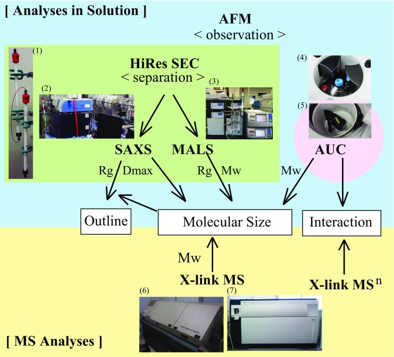
The need for accurate description of protein behavior in solution has gained importance in various fields, including biophysics, biochemistry, structural biology, drug discovery, and antibody drugs. To achieve the desired accuracy, multiple precise analyses should be performed on the target molecule, compared, and effectively combined. This review focuses on the combination of multiple analyses in solution: size-exclusion chromatography (SEC), multi-angle light scattering (MALS), small-angle X-ray scattering (SAXS), analytical ultracentrifugation (AUC), and their complementary methods, such as atomic force microscopy (AFM) and mass spectrometry (MS). We also discuss the comparison between the determined molar mass value of not only the standard proteins, but of a target molecule tubulin and its depolymerizing protein, KIF2, as an example. The comparison of the estimated molar mass value from the different methods provides additional information about the target molecule, because the value reflects the dynamically changing states of the target molecule in solution. The combination and integration of multiple methods will permit a deeper understanding of protein dynamics in solution.

Keywords: Analytical ultracentrifugation (AUC); Kinesin; Microtubule; Multi-angle light scattering (MALS); Size-exclusion chromatography (SEC); Small-angle X-ray scattering (SAXS).


Conflict of interest statement

Conflict of interest

Tadayuki Ogawa declares that he has no conflict of interest. Nobutaka Hirokawa declares that he has no conflict of interest.

Ethical approval

This article does not contain any studies with human participants or animals performed by any of the authors.

Figures

Fig. 1
Fig. 1
High-resolution size-exclusion chromatography (HiRes SEC) displays good separation. a HiRes SEC composed of a tandem connection of twin Superdex 200 Increase 10/300 columns (GE Healthcare). b HiRes SEC displays a good separation of ovalbumin in the monomeric, dimeric, and tetrameric forms. The column was equilibrated with assay buffer (20 mM PIPES, 200 mM KCl, 1 mM MgCl2) and assayed at a flow rate of 0.4 mL/min on an ÄKTA system (GE Healthcare)
Fig. 2
Fig. 2
Combination of HiRes SEC and the refractive index (RI)-multi-angle light scattering (MALS) system. a Protein standard mixture (500 μL) containing ferritin (estimated Mw: 440 kDa), aldolase (158 kDa), ovalbumin (44 kDa), and RNase (13.7 kDa) (100 μg per protein) was separated as mono-disperse fractions by HiRes SEC. Each eluate was directly analyzed by the refractive index (RI) and Dawn Heleos II 18-angle MALS detectors (RI-MALS) (Wyatt). b HiRes SEC-MALS indicated the molecular weight of each protein as 671 kDa for ferritin, 161 kDa for aldolase, 50 KDa for ovalbumin, and 19.8 kDa for RNase. Aggregates at void volume show a large peak. c HiRes SEC-RI detector. The column was equilibrated with assay buffer (20 mM PIPES, 200 mM KCl, 1 mM MgCl2) and assayed at a flow rate of 0.4 mL/min
Fig. 3
Fig. 3
Analytical ultracentrifugation (AUC) determines the molecular mass in solution. a Ovalbumin was analyzed by AUC. b A standard mixture containing ferritin (estimated Mw: 440 kDa), aldolase (158 kDa), ovalbumin (44 kDa), and RNase (13.7 kDa) (100 μg for each protein) was analyzed by AUC
Fig. 4
Fig. 4
Comparison of molar mass values estimated by multiple analyses. a A comparison of the estimated and measured values of the standard proteins: ferritin, aldolase, ovalbumin, and RNase. The molecular weight of the standard proteins was estimated independently by HiRes SEC-MALS and AUC. b Summary of the measured value of tubulin and the KIF2-tubulin 1:2 complex. The molecular weight of tubulin, KIF2-tubulin 1:2 complex was measured by HiRes SEC-MALS, AUC, and crosslink (X-link) matrix-assisted laser desorption/ionization time-of-flight mass spectrometry (MALDI-TOF MS). c The molecular weight of tubulin was analyzed by MALDI-TOF MS (ABI 4700 MALDI-TOF mass spectrometer, SCIEX) in linear mode with a 2,5-dihydroxybenzoic acid (DHB) matrix (WAKO 046-02262)
Fig. 5
Fig. 5
Schematic diagram of the combination of multiple analyses of the target protein. Observation: high-speed atomic force microscopy (HS-AFM) directly monitors the protein sample in solution. Separation: HiRes SEC separates the protein sample and elutes as a mono-disperse fraction. Molecular size determination in solution: based on the separation of HiRes SEC, MALS and SAXS analyses are applied to the mono-disperse sample, to provide accurate measurement of the molecular size in solution. Molecular size determination in vacuo: the peak fraction eluted from HiRes SEC can be analyzed by MALDI-TOF MS to obtain the absolute value. Outline: SAXS analysis provides the outline of the target molecule based on SAXS data and the exact molar mass value. Interaction: the interaction within the complex can be analyzed by X-link MSn. Photos: (1) HiRes SEC composed of a tandem connection of twin Superdex 200 Increase 10/300 columns (GE Healthcare); (2) HiRes SEC-SAXS at beamline BL-15A2 of the Photon Factory of the High Energy Accelerator Research Organization (KEK, Tsukuba, Japan); (3) HPLC (Shimadzu) system equipped with Dawn Heleos II 18-angle MALS detectors (Wyatt); (4) Optima AUC (Beckman Coulter); (5) ProteomeLab XL-A AUC (Beckman Coulter); (6) ABI 4700 MALDI-TOF MS (SCIEX); (7) ultrafleXtreme MALDI-TOF MS (Bruker Daltonics)

Similar articles

Cited by

References

    1. Ando T. Directly watching biomolecules in action by high-speed atomic force microscopy. Biophys Rev. 2017;9:421–429. doi: 10.1007/s12551-017-0281-7. - DOI - PMC - PubMed
    1. Arisaka F, Kanamaru S. Protein interactions in the assembly of the tail of bacteriophage T4. Biophys Rev. 2013;5:79–84. doi: 10.1007/s12551-013-0114-2. - DOI - PMC - PubMed
    1. Arisaka F, Yap ML, Kanamaru S, Rossmann MG. Molecular assembly and structure of the bacteriophage T4 tail. Biophys Rev. 2016;8:385–396. doi: 10.1007/s12551-016-0230-x. - DOI - PMC - PubMed
    1. Desai A, Verma S, Mitchison TJ, Walczak CE. Kin I kinesins are microtubule-destabilizing enzymes. Cell. 1999;96:69–78. doi: 10.1016/S0092-8674(00)80960-5. - DOI - PubMed
    1. Ghirlando R, Mutskova R, Schwartz C. Enrichment and characterization of ferritin for nanomaterial applications. Nanotechnology. 2016;27:045102. doi: 10.1088/0957-4484/27/4/045102. - DOI - PMC - PubMed

LinkOut - more resources