. 2017 Dec 5;12(12):e0189139.
doi: 10.1371/journal.pone.0189139. eCollection 2017.

Z-disc protein CHAPb induces cardiomyopathy and contractile dysfunction in the postnatal heart

Affiliations

Z-disc protein CHAPb induces cardiomyopathy and contractile dysfunction in the postnatal heart

Willemijn van Eldik et al. PLoS One. .

Abstract

Aims: The Z-disc is a crucial structure of the sarcomere and is implicated in mechanosensation/transduction. Dysregulation of Z-disc proteins often result in cardiomyopathy. We have previously shown that the Z-disc protein Cytoskeletal Heart-enriched Actin-associated Protein (CHAP) is essential for cardiac and skeletal muscle development. Furthermore, the CHAP gene has been associated with atrial fibrillation in humans. Here, we studied the misregulated expression of CHAP isoforms in heart disease.

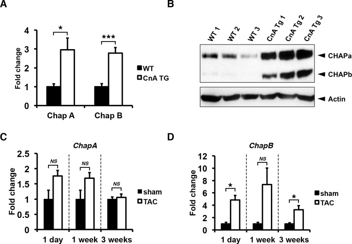
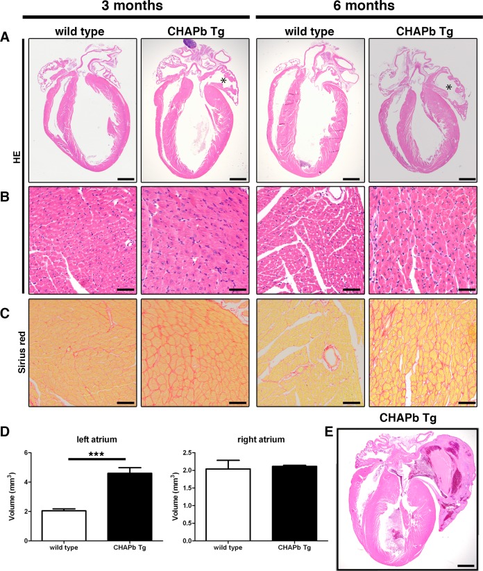
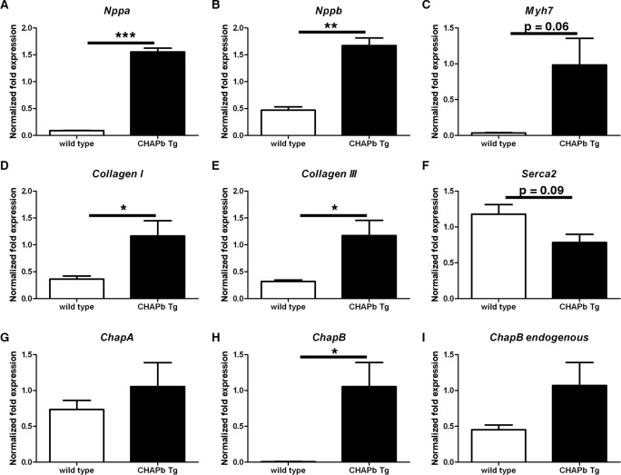
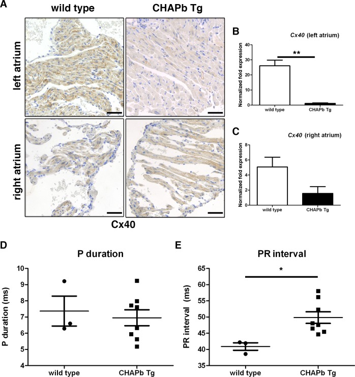
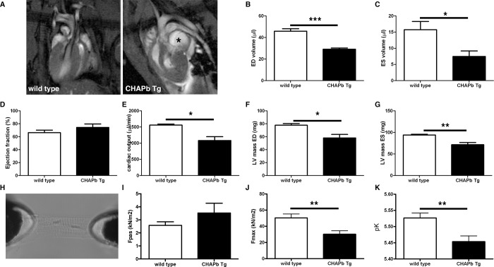
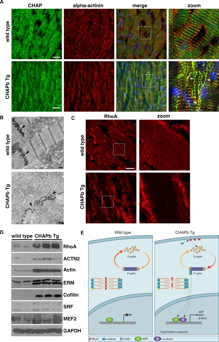
Methods and results: Mice that underwent transverse aortic constriction and calcineurin transgenic (Tg) mice, both models of experimental heart failure, displayed a significant increase in cardiac expression of fetal isoform CHAPb. To investigate whether increased expression of CHAPb postnatally is sufficient to induce cardiomyopathy, we generated CHAPb Tg mice under the control of the cardiac-specific αMHC promoter. CHAPb Tg mice displayed cardiac hypertrophy, interstitial fibrosis and enlargement of the left atrium at three months, which was more pronounced at the age of six months. Hypertrophy and fibrosis were confirmed by evidence of activation of the hypertrophic gene program (Nppa, Nppb, Myh7) and increased collagen expression, respectively. Connexin40 and 43 were downregulated in the left atrium, which was associated with delayed atrioventricular conduction. Tg hearts displayed both systolic and diastolic dysfunction partly caused by impaired sarcomere function evident from a reduced force generating capacity of single cardiomyocytes. This co-incided with activation of the actin signalling pathway leading to the formation of stress fibers.

Conclusion: This study demonstrated that the fetal isoform CHAPb initiates progression towards cardiac hypertrophy, which is accompanied by delayed atrioventricular conduction and diastolic dysfunction. Moreover, CHAP may be a novel therapeutic target or candidate gene for screening in cardiomyopathies and atrial fibrillation.


Conflict of interest statement

Competing Interests: The authors have declared that no competing interests exist.

Figures

Fig 1
Fig 1. Chap is upregulated in mouse models of pathological cardiac hypertrophy.
(A) Quantitative RT-PCR showing increased mRNA levels of both ChapA and B in hearts of transgenic mice expressing constitutively active calcineurin A (CnA Tg) (WT: n = 5, CnA Tg: n = 6; Students t-test: CnA Tg vs WT). (B) Western blot for CHAPa and CHAPb in adult wildtype hearts CnA Tg hearts (WT: n = 5, CnA Tg: n = 6). In wildtype hearts, CHAPa is the dominant isoform while in CnA Tg hearts both CHAPa and b were significantly upregulated. (C) Quantitative RT-PCR for ChapA showing mice subjected to transverse aortic constriction (TAC) for 1 day, 1 week and 3 weeks (n = 4, n = 4, and n = 10 respectively), with no significant regulation of ChapA mRNA compared to sham controls (n = 3, n = 3 and n = 7 respectively). (D) Quantitative RT-PCR for ChapB showing mice subjected to TAC with significant upregulation of ChapB mRNA (number of mice are same as in C). Gapdh was used as internal control in all experiments. T-test with Welch’s correction TAC vs sham: *, p<0.05; **, p<0.01;***, p<0.001.
Fig 2
Fig 2. Hypertrophy and left atrial enlargement in CHAP Tg hearts.
(A-C) Wt (left panels) and CHAPb Tg (right panels) at 3 and 6 months of age. (A) HE stained overview section. The left atrium in CHAPb Tg hearts is enlarged (indicated by *) compared to wt litter mates. (B) Higher magnification of left ventricle. In the left ventricle of CHAPb Tg hearts the cardiomyocytes are hypertrophic. (C) Sirius red staining of the left ventricle showing increase in interstitial fibrosis in CHAPb Tg. (D) Myocardial volume of the left atrium (left panels) and right atrium (right panels) in wt (white bars) and CHAPb Tg (black bars) hearts at 3 months of age (wt n = 4, Tg n = 4, T-test p< 0.001). (E) CHAPb Tg heart with severe phenotype showing pronounced atrial enlargement, filled by a thrombus and thickening of the ventricles. Scale bars 1 mm in A and D, 50 μm in B, C.
Fig 3
Fig 3. Expression of hypertrophy markers and collagens in left ventricle.
qPCR analysis showing mRNA expression of Nppa (A), Nppb (B), Myh7 (C), CollagenI (D) CollagenIII (E), Serca2 (F), ChapA (G), ChapB (H) and endogenous ChapB (I) in the left ventricle of wt (white bars) and CHAPb Tg (black bars) mice. Gapdh, Pgk and H2a were used as internal controls. (wt n = 3, Tg n = 3, T-test: *, p<0.05; **,p<0.01;***, p<0.001).
Fig 4
Fig 4. Decreased Connexin 40 expression in CHAPb Tg left atrium at 6 months of age leads to atrio-ventricular conduction delay.
(A) Immunohistochemical staining showing Connexin 40 expression in wt (left panels) and CHAPb Tg (right panels) left (upper panels) and right (lower panels) atria. (B,C) qPCR analysis of Connexin 40 expression in the left (B) and right (C) atrium (wt n = 3, Tg n = 3 T-test, **,p<0.01). (D,E) ECG analysis of wt (n = 3) and CHAPb Tg (n = 8) mice showing P duration (N.S.) (D) and PR interval (p = 0.02, T-test) (E). Scale bars 50 μm.
Fig 5
Fig 5. CHAPb Tg mice show decrease cardiac performance.
(A) Representative 4-chamber view MRI images of wt and CHAPb Tg heart. Enlarged LA in CHAPb Tg is indicated with *. (B-G) MRI measurements of the left ventricle of wt (n = 4, white bars) and CHAPb Tg (n = 5, black bars) animals at 6 months of age. ED volume (B), ES volume (C), ejection fraction (D), cardiac output (E), LV mass ED (F) and LV mass ES (G). (H) Experimental setup for single membrane-permeabilized cardiomyocyte measurements to determine sarcomere function. Sarcomere force measurements at one month of age showed no difference in passive force (I; Fpas), while maximum force (J; Fmax) and Ca2+-sensitivity (K; pCa50) were reduced in CHAPb Tg (n = 11, black bars) compared to wt (n = 12, white bars). ED (end diastolic), ES (end systolic) and LV (left ventricular). t-test: *, p<0.05; **, p<0.01; ***, p<0.001.
Fig 6
Fig 6. Sarcomeric organization is disturbed and actin signalling is increased in CHAPb Tg hearts.
(A) Immunostaining for CHAP (green) and α-actinin (red) show stress fiber formation (white arrows) in CHAP Tg hearts (B) Electron microscopy analysis of wt and CHAPb Tg hearts at 6 months of age. In CHAPb Tg the sarcomeres were irregular and Z-discs and intercalated discs (black arrow heads) are disorganized, while M-bands are absent (white arrow heads) (C) Wt and CHAPb Tg hearts at 6 months of age stained for RhoA (red). In wt mice RhoA is localized at the membrane of cardiomyocytes and shows a sarcomeric expression pattern. In CHAPb Tg hearts sarcomeric expression of RhoA is absent and membrane expression is increased. (D) Western blot analysis of 2 wt and 3 CHAPb Tg hearts at 6 months of age for RhoA (24kDa), α-actinin (100kDa), actin (42kDa), Ezrin(80 kDa)/moesin(80 kDa)/radixin (75 kDa; ERM), cofilin (19kDa), SRF (40 – 67kDa) and MEF2 (40-65kDa). GAPDH (38kDa) is used as loading control. (E) Working model: in adult wt mice CHAPa is localized at the Z-disc, leading to abundance of monomeric G-actin and subsequent low expression of SRF target genes. In CHAPb Tg mice CHAPb expression results in activation of RhoA, leading to a shift from G-actin to F-actin, binding of co-factors to SRF and activation of SRF target genes, such as ANF, BNP and β-MHC. Scale bars 20 μm in A and C, 1 μm in B.

References

    1. Rohini A, Agrawal N, Koyani CN, Singh R. Molecular targets and regulators of cardiac hypertrophy. Pharmacological research. 2010;61(4):269–80. doi: 10.1016/j.phrs.2009.11.012 - DOI - PubMed
    1. Wilkins BJ, Molkentin JD. Calcium-calcineurin signaling in the regulation of cardiac hypertrophy. Biochemical and biophysical research communications. 2004;322(4):1178–91. doi: 10.1016/j.bbrc.2004.07.121 - DOI - PubMed
    1. Molkentin JD, Lu JR, Antos CL, Markham B, Richardson J, Robbins J, et al. A calcineurin-dependent transcriptional pathway for cardiac hypertrophy. Cell. 1998;93(2):215–28. - PMC - PubMed
    1. Kong SW, Hu YW, Ho JW, Ikeda S, Polster S, John R, et al. Heart failure-associated changes in RNA splicing of sarcomere genes. Circulation Cardiovascular genetics. 2010;3(2):138–46. doi: 10.1161/CIRCGENETICS.109.904698 - DOI - PMC - PubMed
    1. Weeland CJ, van den Hoogenhof MM, Beqqali A, Creemers EE. Insights into alternative splicing of sarcomeric genes in the heart. Journal of molecular and cellular cardiology. 2015;81:107–13. doi: 10.1016/j.yjmcc.2015.02.008 - DOI - PubMed