Morphological study of the eye and adnexa in capuchin monkeys (Sapajus sp.)
- PMID: 29206882
- PMCID: PMC5716594
- DOI: 10.1371/journal.pone.0186569
Morphological study of the eye and adnexa in capuchin monkeys (Sapajus sp.)
Abstract
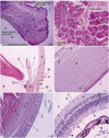
The objective of this study was to describe the anatomic and histologic features of the Sapajus sp. eye, comparing similarities and differences of humans and other species of non-human primates for biomedical research purposes. Computed tomography (CT) of adnexa, eye and orbit live animal, as well as formolized pieces of the same structures of Sapajus sp. for anatomical and histological study were also performed. The anatomical description of the eye and adnexa was performed using the techniques of topographic dissection and exenteration. Histological fragments were fixated in buffered formalin 10%, processed by the routine paraffin inclusion technique, stained with hematoxylin-eosin and special stains. CT scan evaluation showed no differences between the live animal and the formolized head on identification of visual apparatus structures. Anatomic and histologic evaluation revealed rounded orbit, absence of the supraorbital foramen and frontal notch, little exposure of the sclera, with slight pigmentation of the exposed area and marked pigmentation at the sclerocorneal junction. Masson's Trichrome revealed the Meibomian glands, the corneal epithelium and Bowman's membrane; in the choroid, melanocytes and Bruch's membrane were observed; and in the retina, cones and rods as well as, optic nerve, the lamina cribrosa of the nerve fibers bundles. Toluidine blue highlighted the membranes: Bowman, Descemet and the endothelium; in the choroid: melanocytes; and in the retina: nuclear layers and retinal pigment epithelium. In view of the observed results Sapajus sp. is an important experimental model for research in the ophthalmology field, which has been shown due to the high similarity of its anatomical and histological structures with the human species.
Conflict of interest statement
Figures


Similar articles
-
Anatomical, histological and computed tomography comparisons of the eye and adnexa of crab-eating fox (Cerdocyon thous) to domestic dogs.PLoS One. 2019 Oct 23;14(10):e0224245. doi: 10.1371/journal.pone.0224245. eCollection 2019. PLoS One. 2019. PMID: 31644568 Free PMC article.
-
A simplified technique for in situ excision of cornea and evisceration of retinal tissue from human ocular globe.J Vis Exp. 2012 Jun 12;(64):e3765. doi: 10.3791/3765. J Vis Exp. 2012. PMID: 22733120 Free PMC article.
-
Anatomical Study of Intrahemispheric Association Fibers in the Brains of Capuchin Monkeys (Sapajus sp.).Biomed Res Int. 2015;2015:648128. doi: 10.1155/2015/648128. Epub 2015 Nov 29. Biomed Res Int. 2015. PMID: 26693488 Free PMC article.
-
Advances in myopia research anatomical findings in highly myopic eyes.Eye Vis (Lond). 2020 Sep 2;7:45. doi: 10.1186/s40662-020-00210-6. eCollection 2020. Eye Vis (Lond). 2020. PMID: 32905133 Free PMC article. Review.
-
Anointing variation across wild capuchin populations: a review of material preferences, bout frequency and anointing sociality in Cebus and Sapajus.Am J Primatol. 2012 Apr;74(4):299-314. doi: 10.1002/ajp.20971. Epub 2011 Jul 18. Am J Primatol. 2012. PMID: 21769906 Review.
Cited by
-
Anatomical, histological and computed tomography comparisons of the eye and adnexa of crab-eating fox (Cerdocyon thous) to domestic dogs.PLoS One. 2019 Oct 23;14(10):e0224245. doi: 10.1371/journal.pone.0224245. eCollection 2019. PLoS One. 2019. PMID: 31644568 Free PMC article.
References
-
- Bicca-Marques JC, Gomes DF. Birth seasonality of Cebus apella (Platyrrhini, Cebidae) in brazilian zoos along a latitudinal gradient. Am J Primatol. 2005; 65: 141–147. doi: 10.1002/ajp.20104 . - DOI - PubMed
-
- Martin RD. Primates. Current Biology. 2012; 22 (18): 785–790. http://dx.doi.org.10.1016/j.cub.2012.07.015 - PubMed
-
- Verona CE, Pissinatti A. Primates–Primatas do Novo Mundo (Sagui, Macaco-prego, Macaco-aranha, Bugio e Muriqui) In: Cubas ZS, Silva JCR, Catão-Dias JL, editors. Tratado de Animais Selvagens Medicina Veterinária. 2nd ed São Paulo: Roca; 2014. p. 723–743.
-
- Sasaki E, Suemizu H, Shimada A, Hanazawa K, Oiwa R, Kamioka M, et al. Generation of transgenic non-human primates with germline transmission. Nature. 2009; 459: 523–527. doi: 10.1038/nature08090 - DOI - PubMed
-
- Torres LB, Araújo BHS, Castro PHG, Cabral FR, Marruaz KS, Araújo MS. The Use of New World Primates for Biomedical Research: An Overview of the Last Four Decades. Am J Primatol. 2010; 72: 1055–1061. doi: 10.1002/ajp.20864 . - DOI - PubMed
MeSH terms
LinkOut - more resources
Full Text Sources
Other Literature Sources
Medical

