. 2018 Feb 1;28(2):61-68.
doi: 10.1093/glycob/cwx098.

Tunicamycin-induced ER stress in breast cancer cells neither expresses GRP78 on the surface nor secretes it into the media

Affiliations

Tunicamycin-induced ER stress in breast cancer cells neither expresses GRP78 on the surface nor secretes it into the media

Jesús E Serrano-Negrón et al. Glycobiology. .

Erratum in

Abstract

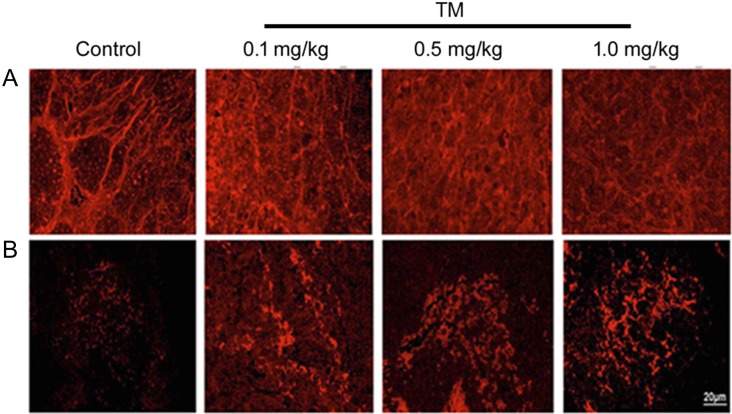
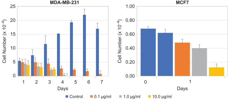
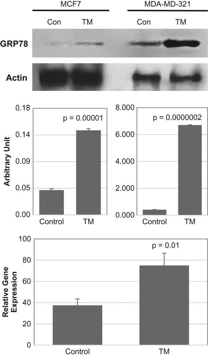
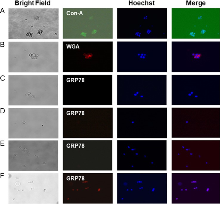
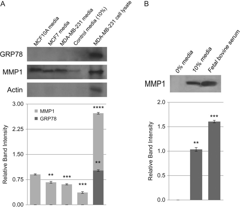
GRP78 (an Mr 78 kDa calcium dependent glucose binding protein) is located in ER lumen. It functions as ER chaperone and translocates proteins for glycosylation at the asparagine residue present in the sequon Asn-X-Ser/Thr. Paraffin sections from N-glycosylation inhibitor tunicamycin treated ER-/PR-/HER2+ (double negative) breast tumor in athymic nude mice exhibited reduced N-glycan but increased GRP78 expression. We have evaluated the effect of tunicamycin on cellular localization of GRP78 in metastatic human breast cancer cells MDA-MB-231 (ER-/PR-/HER2-). Tunicamycin inhibited cell proliferation in a time and dose-dependent manner. Nonmetastatic estrogen receptor positive (ER+) MCF-7 breast cancer cells were also equally effective. GRP78 expression (protein and mRNA) was higher in tunicamycin (1.0 μg/mL) treated MCF-7 and MDA-MB-231 cells. GRP78 is an ER stress marker, so we have followed its intracellular localization using immunofluorescence microscopy after subjecting the cancer cells to various stress conditions. Unfixed cells stained with either FITC-conjugated Concanavalin A (Con A) or Texas-red conjugated wheat germ agglutinin (WGA) exhibited surface expression of N-glycans but not GRP78. GRP78 became detectable only after a brief exposure of cells to ice-cold methanol. Western blotting did not detect GRP78 in conditioned media of cancer cells whereas it did for MMP-1. The conclusion, GRP78 is expressed neither on the outer-leaflet of the (ER-/PR-/HER2-) human breast cancer cells nor it is secreted into the culture media during tunicamycin-induced ER stress. Our study therefore suggests strongly that anti-tumorigenic action of tunicamycin can be modeled to develop next generation cancer therapy, i.e., glycotherapy for treating breast and other sold tumors.

Keywords: ER stress; GRP78; asparagine-linked glycoprotein; breast cancer; tunicamycin.


Figures

Fig. 1.
Fig. 1.
Expression of cell surface glycan and induction of ER stress in ER/PR/HER2+ breast tumor. Orthotopic double negative (ER/PR/HER2+) breast tumor was developed in athymic nude mice with MDA-MB-435 human breast cancer cells. Formalin-fixed and paraffin-embedded sections were processed as before [Quinoñes et al. 2008; Banerjee et al. 2011]. (A) WGA staining for N-glycan in control and tunicamycin (TM) (0–1.0 mg/kg)-treated breast tumor tissue sections. Images were captured under a fluorescence microscope. (B) Immunocytochemistry of GRP78 in paraffin section of breast tumor tissue. The sections were stained with anti-GRP78 antibody followed by rhodamine-conjugated secondary antibody. Histology scale: 20 μm.
Fig. 2.
Fig. 2.
Tunicamycin inhibits proliferation of ER/PR/HER2 (triple negative; MDA-MB-231) and estrogen receptor positive (MCF-7) breast cancer cells. Triple negative human breast cancer cells (MDA-MB-231) were seeded (2 × 10⁴ cells/well) in 24-well plates, synchronized in serum-free media for 24 h and treated with tunicamycin (0, 0.1, 1.0 and 10.0 μg/mL) in a 2% serum-containing media for 7 days. The cells were counted after every 24 h in a haemocytometer. Estrogen receptor positive human breast cancer cells (MCF-7) were seeded (2 × 10⁴ cells/well) in 24-well plates, synchronized in serum-free media for 48 h and treated with tunicamycin (0, 0.1, 1.0 and 10.0 μg/mL) in a 2% serum-containing media for 24 h. The cells were counted in a haemocytometer. The error bars represent the standard deviations (mean ± SD) for n = 3.
Fig. 3.
Fig. 3.
GRP78 Protein and mRNA expression in ER+ (estrogen receptor positive) and ER/PR/HER2 (triple negative; MDA-MB-231) human breast cancer cells. Triple negative MDA-MB-231 and ER+ MCF-7 breast cancer cells were cultured, synchronized and treated with tunicamycin (1 μg/mL) for 48 h. Top: GRP78 protein expression was examined by immunoblotting (30 μg of total protein) from control (Con) and Tunicamycin (TM) treated cells. The blots were developed with anti-GRP78 antibody (1:1000; v/v) and anti-actin antibody (1:5000; v/v). Middle: The histogram representing the quantification of GRP78 expression by Image J software, respectively. The results are an average from three blots done independently. Bottom: Quantification of GRP78 mRNA expression in triple negative breast cancer cells before and after tunicamycin treatment. The error bars represent the standard error (mean ± SE) for n = 3, and the P-values are from Student t-test.
Fig. 4.
Fig. 4.
Localization of GRP78 in ER/PR/HER2 (triple negative; MBA-MB-231) breast cancer cells. Cells were cultured overnight, removed by nonenzymatic cell dissociation solution. The surface N-glycans were stained with (A) FITC-conjugated Con-A (green, 20×), and (B) Texas-red conjugated WGA (red; 20×) in a 50 mM Tris buffer, pH 7.0 containing 0.15 M NaCl and 4 mM CaCl2 but no fixative. The images were collected by a fluorescence microscope (Axioskop 2, Carl Zeiss, Germany) with AxioCam MRc5 camera and Axion Vision Rel 4.6 software. (C–E) Detection of GRP78 (red, 40×); (C) cells were cultured for 24 h in EMEM with 10% serum and then stained with anti-GRP78 antibody; (D) cells were cultured for 24 h in EMEM without serum (i.e., serum-free; 0% FBS) and then stained with anti-GRP78 antibody; (E) cells were treated with tunicamycin (1 μg/mL) for 24 h in EMEM containing 2% serum and the stained with anti-GRP78 antibody; (F) cells were treated with tunicamycin (1 μg/mL) as in (E) and then permeabilized by exposing to ice-cold methanol for 15 s and stained with anti-GRP78 antibody.
Fig. 5.
Fig. 5.
Secretion of GRP78 and MMP-1 in conditioned media of breast cancer cells. Breast cancer cells MCF-7 and MDA-MB-231 as well as the control MCF-10A were all cultured overnight in EMEM containing 10% fetal bovine serum. The conditioned media were collected, dialyzed overnight at 4°C in 50 mM Tris-HCl, pH 7.0, and concentrated approximately 1000-fold over Aquacide II™. Equal amount of protein from each sample was analyzed by western blotting. (A) GRP78; lane 1 = media from MCF-10A cells, lane 2 = media from MCF-7 cells, lane 3 = media from MDA-MB-231 cells, lane 4 = EMEM with 10% serum, lane 5 = MDA-MB-231 whole cell lysate; (B) MMP-1; lane 1 = EMEM with 0% fetal bovine serum, lane 2 = EMEM with 10% fetal bovine serum, lane 3 = fetal bovine serum from stock. The error bars represent the standard error (Mean ± SE) for n = 3. P-values are from the Student t-test (** = P < 0.005; *** = P > 0.00005; **** = P > 0.000005).

References

    1. Alberts B, Johnson A, Lewis J, Raff M, Roberts K, Peter Walter P. 2008. Molecular Biology of the Cell, 5th ed New York: Garland Science.
    1. Arap MA, Lahdenranta J, Mintz PJ, Hajitou A, Sarkis AS, Arap W, Pasqualini R. 2004. Cell surface expression of the stress response chaperone GRP78 enables tumor targeting by circulating ligands. Cancer Cell. 6:275–284. - PubMed
    1. Banerjee A, Lang JY, Hung MC, Sengupta K, Banerjee SK, Baksi K, Banerjee DK. 2011. Unfolded protein response is required in nu/nu mice microvasculature for treating breast tumor with tunicamycin. J Biol Chem. 286:29127–29138. - PMC - PubMed
    1. Banerjee DK, Ornberg RL, Youdim MB, Heldman E, Pollard HB. 1985. Endothelial cells from Bovine adrenal medulla develop capillary-like growth patterns in culture. Proc Natl Acad Sci USA. 82:4702–4706. - PMC - PubMed
    1. Chiu CC, Lin CY, Lee LY, Chen YJ, Kuo TF, Chang JT, Liao CT, Wang HM, Yen TC, Shen CR et al. . 2008. Glucose-regulated protein 78 regulates multiple malignant phenotypes in head and neck cancer and may serve as a molecular target of therapeutic intervention. Mol Cancer Ther. 7:2788–2797. - PubMed

Publication types