. 2018 Feb;41(2):749-756.
doi: 10.3892/ijmm.2017.3283. Epub 2017 Nov 24.

Effects of kartogenin on the attenuated nucleus pulposus cell degeneration of intervertebral discs induced by interleukin-1β and tumor necrosis factor-α

Affiliations

Effects of kartogenin on the attenuated nucleus pulposus cell degeneration of intervertebral discs induced by interleukin-1β and tumor necrosis factor-α

Yao Huang et al. Int J Mol Med. 2018 Feb.

Abstract

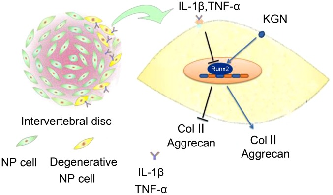
Cytokines are the main cause of intervertebral disc degeneration. Kartogenin (KGN) is found to protect chondrocytes from cytokines. To explore whether KGN can slow down the degeneration on intervertebral discs following exposure to interleukin-1β (IL-1β) and tumor necrosis factor-α (TNF‑α), the expression of type II collagen (Col II) and aggrecan were detected by immunofluorescence, immunohistochemistry and tissue staining. An in vitro model of disc degeneration using human nucleus pulposus cells (hNPCs) and ex vivo culture of mouse intervertebral discs organs under the actions of inflammatory cytokines were used, and the expression of Col II and aggrecan in hNPCs were detected by semi-quantitative western blot analysis, and the mRNA expression of the genes than encode Col II and aggrecan were detected by reverse transcription‑quantitative polymerase chain reaction (RT-qPCR). The results indicated that the expression of Col II and aggrecan was reduced in the degeneration models. However, the protein expressions of Col II and aggrecan were significantly elevated in hNPCs and the mouse intervertebral discs following addition of KGN. RT-qPCR results revealed that the mRNA expression of Col II and aggrecan was increased in hNPCs and mouse intervertebral discs following treatment with KGN. Thus, KGN effectively increased the expression of Col II and aggrecan in hNPCs and slowed the degeneration of intervertebral discs stimulated by IL-1β and TNF-α.


Figures

Figure 1
Figure 1
Col II and aggrecan expression were elevated by KGN in hNPCs. Expression of Col II and aggrecan in hNPCs after 24 and 48 h by immunofluorescence. (A) Col II, 24 h; (B) Col II, 48 h; (C) aggrecan, 24 h; and (D) aggrecan, 48 h. The expression of Col II and aggrecan were significantly inhibited in the group under inflammatory cytokines alone. However, the expression of Col II and aggrecan were elevated by KGN (original magnification, ×200). Col II, type II collagen; KGN, kartogenin; hNPC, human nucleus pulposus cells; IL-1β, interleukin-1β; TNF-α, tumor necrosis factor-α; C, control; I+T, IL-1β + TNF-α; I+T+KL, IL-1β + TNF-α + low concentration of KGN; I+T+KH, IL-1β + TNF-α + high concentration of KGN.
Figure 2
Figure 2
KGN promoted the expression of Col II and aggrecan in mouse intervertebral discs. (A) Immunohistochemisty of Col II and (B) aggrecan staining in paraffin sections of mouse intervertebral discs (at 3 and 10 days). Col II expression in the group treated with KGN was higher than that in the group under inflammatory cytokines. The group under inflammatory cytokines had a collapsed nucleus pulposus, thereby indicating the extracellular matrix loss in the intervertebral disc. However, the group with KGN did not exhibit such a phenomenon, with less loss of the extracellular matrix from the nucleus pulposus tissues in the intervertebral discs (original magnification, ×40). Col II, type II collagen; KGN, kartogenin; IL-1β, interleukin-1β; TNF-α, tumor necrosis factor-α; C, control; I+T, IL-1β + TNF-α; I+T+KL, IL-1β + TNF-α + low concentration of KGN; I+T+KH, IL-1β + TNF-α + high concentration of KGN.
Figure 3
Figure 3
KGN promoted the release of Col II and aggrecan. (A) Western blot analysis of Col II and aggrecan expression in hNPCs (at 24 and 48 h). (B) Expression of Col II and aggrecan in the nucleus pulposus of mouse intervertebral discs by western blot analysis (at 3 and 10 days). The expression of Col II and aggrecan in the groups with KGN (especially at a high concentration) were higher than those in the group under inflammatory cytokines but did not reach the levels of the blank control group. (C) Densitometry of western blot analysis showed KGN improved the expression of Col II and aggrecan in hNPCs and intervertebral discs. Data presented as mean ± standard deviation of three independent experiments performed in triplicate (n=3). *P<0.05 and **P<0.01. Col II, type II collagen; KGN, kartogenin; hNPC, human nucleus pulposus cells; IL-1β, interleukin-1β; TNF-α, tumor necrosis factor-α; C, control; I+T, IL-1β + TNF-α; I+T+KL, IL-1β + TNF-α + low concentration of KGN; I+T+KH, IL-1β + TNF-α + high concentration of KGN.
Figure 4
Figure 4
KGN increased the mRNA expression of Col II and aggrecan. Reverse transcription-quantitative polymerase chain reaction of genes associated with Col II and aggrecan in (A) human nucleus pulposus cells and (B) mouse intervertebral discs. The inflammatory cytokines significantly inhibited the expression of genes associated with Col II and aggrecan. Their expression was elevated by adding KGN; although their expression levels did not reach that of the blank control group, the difference was still statistically significant compared with the group under inflammatory cytokines (*P<0.05 and **P<0.001). Col II, type II collagen; col2a1, collagen type II α 1 chain; AGC1, aggrecan; KGN, kartogenin; IL-1β, interleukin-1β; TNF-α, tumor necrosis factor-α; C, control; I+T, IL-1β + TNF-α; I+T+KL, IL-1β + TNF-α + low concentration of KGN; I+T+KH, IL-1β + TNF-α + high concentration of KGN.
Figure 5
Figure 5
RUNX2 expression was elevated by KGN. RUNX2 expression in human nucleus pulposus cells and mouse intervertebral discs were inhibited by inflammatory cytokines. However, it was elevated after the treatment of KGN, especially at a high concentration (*P<0.05 and **P<0.001). RUNX2, runt related transcription factor 2; KGN, kartogenin; IL-1β, interleukin-1β; TNF-α, tumor necrosis factor-α; C, control; I+T, IL-1β + TNF-α; I+T+KL, IL-1β + TNF-α + low concentration of KGN; I+T+KH, IL-1β + TNF-α + high concentration of KGN.
Figure 6
Figure 6
IL-1β and TNF-α improved the degeneration of NP cells and discs by interrupt the expression of RUNX2. KGN effectively promoted the expression of RUNX2, thus increased the Col II and aggrecan, and rivaled to the degeneration of disc induced by cytokines. KGN effectively delayed the degeneration of intervertebral disc. IL-1β, interleukin-1β; TNF-α, tumor necrosis factor-α; KGN, kartogenin; Col II, type II collagen; NP, nucleus pulposus; RUNX2, runt related transcription factor 2.

Similar articles

Cited by

References

    1. Andersson GB. Epidemiological features of chronic low-back pain. Lancet. 1999;354:581–585. doi: 10.1016/S0140-6736(99)01312-4. - DOI - PubMed
    1. Katz JN. Lumbar disc disorders and low-back pain: socioeconomic factors and consequences. J Bone Joint Surg A. 2006;88(Suppl 2):21–24. - PubMed
    1. Jenkins H. Classification of low back pain. Australas Chiropr Osteopathy. 2002;10:91–97. - PMC - PubMed
    1. Le Maitre CL, Freemont AJ, Hoyland JA. The role of interleukin-1 in the pathogenesis of human intervertebral disc degeneration. Arthritis Res Ther. 2005;7:R732–R745. doi: 10.1186/ar1732. - DOI - PMC - PubMed
    1. Séguin CA, Pilliar RM, Roughley PJ, Kandel RA. Tumor necrosis factor-alpha modulates matrix production and catabolism in nucleus pulposus tissue. Spine. 2005;30:1940–1948. doi: 10.1097/01.brs.0000176188.40263.f9. - DOI - PubMed

MeSH terms