. 2018 Feb;41(2):800-808.
doi: 10.3892/ijmm.2017.3270. Epub 2017 Nov 17.

Autophagy relieves the function inhibition and apoptosis‑promoting effects on osteoblast induced by glucocorticoid

Affiliations

Autophagy relieves the function inhibition and apoptosis‑promoting effects on osteoblast induced by glucocorticoid

Yudi Han et al. Int J Mol Med. 2018 Feb.

Abstract

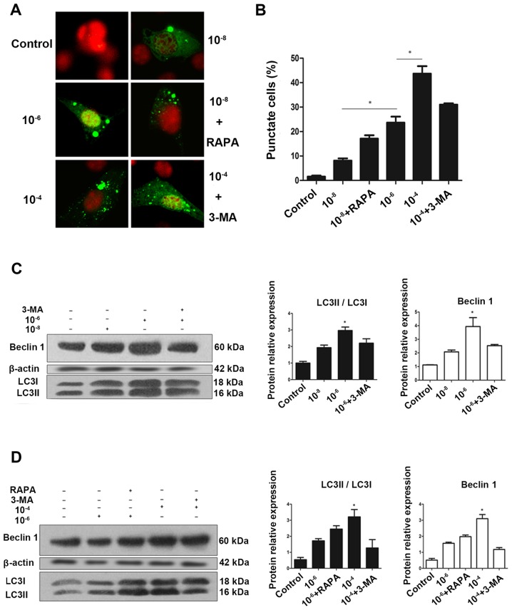
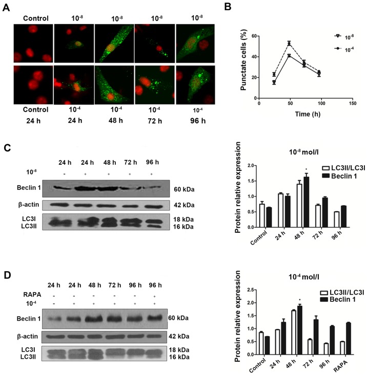
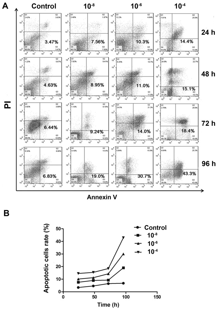
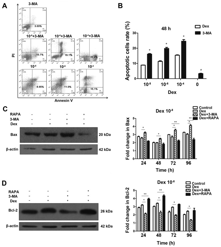
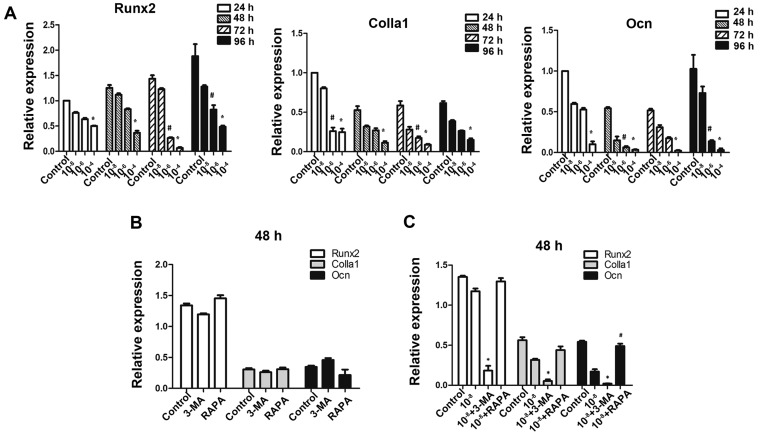
Autophagy may be a major mechanism by which osteoblasts (OBs) protect against the negative effects of chronic glucocorticoid (GC) usage. OBs are closely associated with the remodeling that occurs in GC‑induced osteoporosis (GIO). In osteocytes, in response to stress induced by GCs, several pathways are activated, including cell necrosis, apoptosis and autophagy. However, the role of autophagy in OBs following treatment with excess GCs has not been addressed. In the current study, confocal microscopy observation of green fluorescent protein‑microtubule‑associated protein 1 light chain 3β (LC3) punctuate, and western blotting for LC3Ⅱ and Beclin 1 were performed for detection of autophagy in the MC3T3‑E1 osteoblastic cell line. Flow cytometry and western blotting were used for the examination of apoptosis and expression of BAX apoptosis regulator (Bax)/apoptosis regulator Bcl‑2 (Bcl‑2). The expression of genes associated with osteoblastic function, runt‑related transcription factor 2, α‑1 type 1 collagen and osteocalcin, were measured by reverse transcription‑quantitative polymerase chain reaction. The results indicated that autophagy was induced in OBs during dexamethasone (Dex) treatment in a dose‑dependent manner. The level of autophagy did not continue to increase over time, but peaked at 48 h and then decreased gradually. Subsequently, flow cytometry was used to demonstrate that inhibition of autophagy induced apoptosis in OBs under Dex treatment, and was associated with the upregulation of Bax and the downregulation of Bcl‑2 protein expression. Furthermore, the data suggested that the inhibition of autophagy also suppressed the expression of osteoblastic genes. By contrast, the stimulation of autophagy maintained the gene expression level under Dex treatment. The data revealed that autophagy is an important regulator of osteoblastic apoptosis through its interaction with Bax/Bcl‑2, and maintains the osteoblastic function of MC3T3‑E1 cells following GC exposure. In addition, these results indicated that the suppression of autophagy in OBs under chronic GC therapy may increase the prevalence of GIO and fragility fractures.


Figures

Figure 1
Figure 1
Autophagy is activated during glucocorticoid treatment. (A) Cells transiently transfected plasmid expressing eGFP-LC3B fusion protein, were exposed to Dex or control (no Dex) for 24 h. Representative fluorescence microscopy images are presented (magnification, ×100). (B) Control group had <2% of cells expressing punctate eGFP-LC3. Whereas, 24 h treatment with 10−8, 10−6 and 10−4 mol/l Dex led to eGFP-LC3 puncta formation in ~50% of cells. *P<0.05 vs. control, 10−8, and 10−6 + 3-MA groups. Cells were exposed to culture medium containing 10−8, 10−6 and 10−4 mol/l Dex for 24 h with (C) 3-MA (400 μmol/l) added 1.5 h before the experiment plus (D) RAPA (500 nmol/l) added during the last 6 h of the experiment. Autophagy markers were evaluated by western blotting. Positive blots (right) and corresponding semiquantitative analysis (left) are presented. Values are presented as the mean ± standard deviation (n=3). *P<0.05 vs. control, 10−6, 10−6 + RAPA, and 10−4 + 3-MA groups. Dex, dexamethasone; eGFP, enhanced green fluorescent protein; RAPA, rapamycin; 3-MA, 3-methyladenine; LC3, microtubule-associated protein 1 light chain 3β.
Figure 2
Figure 2
Generation and characterization of autophagy over time. (A) Confocal microscopy images of MC3T3-E1 cells treated with 10−8 and 10−4 mol/l Dex for 24, 48, 72 and 96 h (magnification, ×100). (B) Broken line graph showed that enhanced green fluorescent protein-LC3 puncta positive cells reached a peak at 48 h and then decreased. Representative immunoblots and densitometry analysis of cells treated with (C) Dex at 10−8 or (D) Dex at 10−4 mol/l for 24 to 96 h and RAPA (500 nmol/l) for 96 h. Corresponding densitometric quantification showed that autophagy markers reached a peak at 48 h and decrease gradually after that. *P<0.05 vs. other time groups. Dex, dexamethasone; LC3, microtubule-associated protein 1 light chain 3β; RAPA, rapamycin.
Figure 3
Figure 3
Apoptosis is induced during Dex treatment. (A) Annexin V-fluorescein isothiocyanate/PI staining was analyzed by flow cytometry. Representative dot plots of MC3T3-E1 cells subjected to 10−8, 10−6 and 10−4 mol/l Dex or control are presented with the percentage of Annexin V-positive cells indicated. (B) Population of apoptotic cells was increased in a dose-dependent manner. There was significant difference between 10−6 mol/l and control group (P<0.05), 10−4 mol/l and control group (P<0.05) at all time-points. Dex, dexamethasone; PI, propidium iodide.
Figure 4
Figure 4
Regulation of autophagy interferes in the negative effects of Dex on MC3T3-E1 cells. (A) MC3T3-E1 cells were treated with Dex, in the presence or absence of chemical inhibitors of autophagy 3-MA. Annexin V-fluorescein isothiocyanate/PI staining was analyzed by flow cytometry. Representative dot plots of MC3T3-E1 cells subjected to Dex or 3-MA alone are presented. (B) Data expressed as percentage of Annexin V-positive cells (n=3). *P<0.05, vs. corresponding control and #P<0.05 other groups vs. 3-MA alone. Cells were treated with 10−8 mol/l Dex and 3-MA (400 μmol/l) or RAPA (500 nmol/l), and the expression of (C) Bax and (D) Bcl-2 were detected by western blotting. The results were representative of three independent experiments. β-actin was used as a loading control. *P<0.05; **P<0.05. 3-MA, 3-methyladenine; PI, propidium iodide; Dex, dexamethasone; RAPA, rapamycin; Bax, BAX apoptosis regulator; Bcl-2, Bcl-2 apoptosis regulator.
Figure 5
Figure 5
Autophagy has a protective effect on osteoblastic gene expressions of MC3T3-E1 cells under Dex treatment. MC3T3-E1 cells were treated with various doses of Dex. (A) Osteogenesis-associated genes Runx2, Colla1 and Ocn were examined by reverse transcription-quantitative polymerase chain reaction. *P<0.05 vs. control; #P<0.05 vs. control. (B) Cells treated with 3-MA (400 μmol/l) or RAPA (500 nmol/l) alone were also examined, no significant difference was detected (P>0.05). (C) Cells were co-treated with Dex and 3-MA or RAPA. *P<0.05 vs. Dex group; #P<0.05 vs. Dex group. Dex, dexamethasone; Runx2, runt-related transcription factor 2; Colla1, α-1 type 1 collagen; Ocn, osteocalcin; 3-MA, 3-methyladenine; RAPA, rapamycin.

References

    1. Kanis JA, Johansson H, Oden A, Johnell O, de Laet C, Melton LJ, III, Tenenhouse A, Reeve J, Silman AJ, Pols HA, et al. A meta-analysis of prior corticosteroid use and fracture risk. J Bone Miner Res. 2004;19:893–899. doi: 10.1359/JBMR.040134. - DOI - PubMed
    1. Van Staa TP, Laan RF, Barton IP, Cohen S, Reid DM, Cooper C. Bone density threshold and other predictors of vertebral fracture in patients receiving oral glucocorticoid therapy. Arthritis Rheum. 2003;48:3224–3229. doi: 10.1002/art.11283. - DOI - PubMed
    1. Weinstein RS. Clinical practice. Glucocorticoid-induced bone disease. N Engl J Med. 2011;365:62–70. doi: 10.1056/NEJMcp1012926. - DOI - PubMed
    1. Mankin HJ. Nontraumatic necrosis of bone (osteonecrosis). N Engl J Med. 1992;326:1473–1479. doi: 10.1056/NEJM199205283262206. - DOI - PubMed
    1. LoCascio V, Bonucci E, Imbimbo B, Ballanti P, Adami S, Milani S, Tartarotti D, DellaRocca C. Bone loss in response to long-term glucocorticoid therapy. Bone Miner. 1990;8:39–51. doi: 10.1016/0169-6009(91)90139-Q. - DOI - PubMed

MeSH terms