. 2018 Feb;41(2):908-914.
doi: 10.3892/ijmm.2017.3290. Epub 2017 Nov 27.

Pentosan polysulfate ameliorates apoptosis and inflammation by suppressing activation of the p38 MAPK pathway in high glucose‑treated HK‑2 cells

Affiliations

Pentosan polysulfate ameliorates apoptosis and inflammation by suppressing activation of the p38 MAPK pathway in high glucose‑treated HK‑2 cells

Ping Chen et al. Int J Mol Med. 2018 Feb.

Abstract

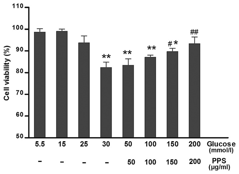
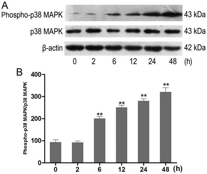
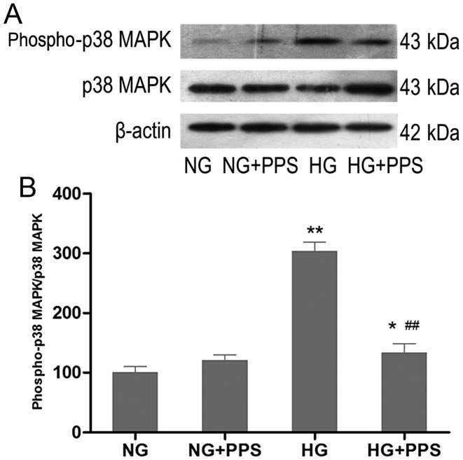
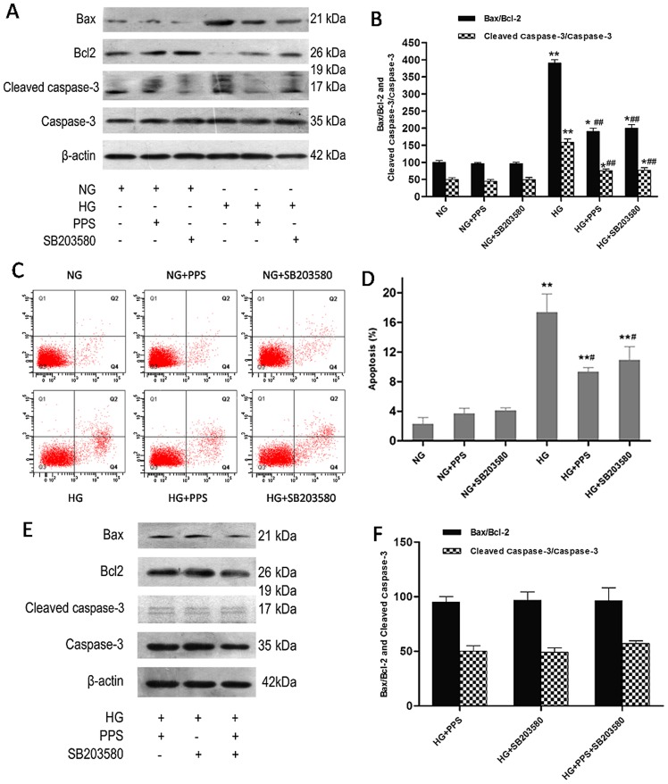
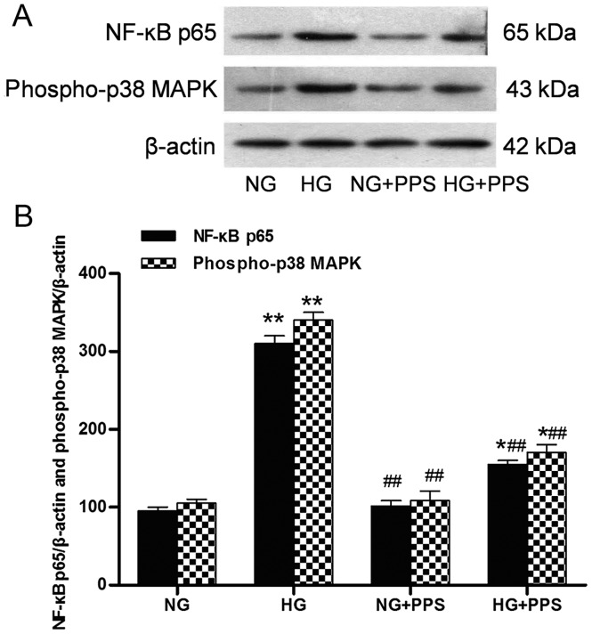
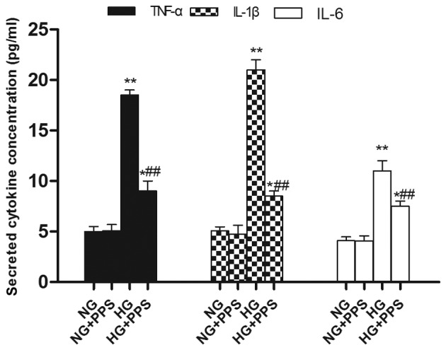
The apoptosis of tubular epithelial cells in diabetic nephropathy (DN) is commonly observed in human renal biopsies. Inflammation plays a key role in DN, and pentosan polysulfate (PPS) has been shown to largely attenuate the inflammation of nephropathy in aging diabetic mice. p38 mitogen‑activated protein kinase (p38 MAPK) plays a crucial role in tissue inflammation and cell apoptosis, and it is activated by hyperglycemia. In the present study, high glucose (HG)‑treated human renal proximal tubular epithelial cells (HK‑2) were used to examine the protective effects of PPS against HG‑stimulated apoptosis and inflammation. The results of the study revealed that PPS markedly suppressed the HG‑induced reduction in cell viability. Incubation of HK‑2 cells with HG activated the p38 MAPK pathway and, subsequently, as confirmed by western blot analysis and flow cytometry, increased cell apoptosis, which was blocked by PPS. In addition, PPS treatment significantly inhibited HG‑stimulated p38 MAPK and nuclear factor‑κB activation, and reduced the production of pro‑inflammatory cytokines, such as tumor necrosis factor‑α, interleukin (IL)‑1β and IL‑6. In conclusion, PPS ameliorates p38 MAPK‑mediated renal cell apoptosis and inflammation. The anti‑apoptotic actions and anti‑inflammatory effects of PPS prompt further investigation of this compound as a promising therapeutic agent against DN.


Figures

Figure 1
Figure 1
Cell viability. HK-2 cells were treated with 50–200 µg/ml PPS and assessed using the MTT assay. Values are presented as means ± standard deviation of at least three determinations. *P<0.05, **P<0.01 compared with 5 mmol/l glucose treatment. #P<0.05, ##P<0.01 compared with 30 mmol/l glucose treatment. PPS, pentosan polysulfate.
Figure 2
Figure 2
Effect of HG treatment on the expression of phospho-p38 MAPK in HK-2 cells. (A) The expression levels of p38 MAPK and phospho-p38 MAPK in cultured HK-2 cells exposed to HG (30 mmol/l) were examined using western blot analysis. Representative western blots of phospho-p38 MAPK in experimental groups at various time-points (n=6). (B) Densitometric analysis of western blots. Data are presented as the means from triplicate determinations (means ± standard deviation) indicated by the vertical bars (**P<0.01 vs. control). HG, high glucose; p38 MAPK, p38 mitogen-activated protein kinase.
Figure 3
Figure 3
Effect of PPS on the expression of HG-stimulated phospho-p38 MAPK in HK-2 cells. (A) HK-2 cells were grown in HG and NG in the absence or presence of PPS for 48 h. Phospho-p38 MAPK expression was determined by western blot analysis. (B) Densitometric scan analysis for western blotting. NG: 5.5 mmol/l D-glucose; NG + PPS: 5.5 mmol/l D-glucose plus 200 µg̸ml PPS; HG: 30 mmol/l D-glucose; and HG + PPS: 30 mmol/l D-glucose plus 200 µg/ml PPS. Data are presented as the means from triplicate determinations (means ± standard deviation) indicated by the vertical bars (*P<0.05, **P<0.01 vs. NG and ##P<0.01 vs. HG). PPS, pentosan polysulfate; HG, high glucose; NG, normal glucose; p38 MAPK, p38 mitogen-activated protein kinase.
Figure 4
Figure 4
Effect of PPS or SB2035 on HG-induced apoptosis in HK-2 cells. (A) HK-2 cells were grown in HG and NG in the absence or presence of PPS or SB203580 for 48 h. The expression levels of caspase-3, cleaved caspase-3, Bax, Bcl-2 and β-actin were determined using western blot analysis. (B) Densitometric analysis for western blot analysis. (C) Apoptosis of HK-2 cells was determined by flow cytometry. (D) Analysis of apoptotic rate using flow cytometry. (E) Effects of PPS and SB203580 on the expression of caspase-3, cleaved caspase-3, Bax, Bcl-2 and β-actin were determined using western blot analysis. (F) Densitometric analysis for western blotting. NG: 5.5 mmol/l D-glucose; NG + PPS: 5.5 mmol/l D-glucose plus 200 µg/ml PPS; NG + SB203580: 5.5 mmol/l D-glucose plus 10 µM SB203580; HG: 30 mmol/l D-glucose; HG + PPS: 30 mmol/l D-glucose plus 200 µg/ml PPS; HG + SB203580: 30 mmol/l D-glucose plus 10 µM SB203580; and HG + PPS + SB203580: 30 mmol/l M D-glucose plus 200 µg/ml PPS plus 10 µM SB203580. Experiments were repeated at least three times, and the results are presented as the mean ± standard deviation. *P<0.05 and **P<0.01 vs. NG; #P<0.01 vs. HG; ##P<0.01 vs. HG. PPS, pentosan polysulfate; HG, high glucose; NG, normal glucose.
Figure 5
Figure 5
Effects of PPS on phospho-p38 MAPK and NF-κB p65 expression in cultured HK-2 cells. (A) Western blot analysis of phospho-p38 MAPK and NF-κB p65 activities in HK-2 cells. (B) Densitometric data of protein analysis. NG: 5.5 mmol/l D-glucose; HG: 30 mmol/l D-glucose; NG + PPS: 5.5 mmol/l D-glucose plus 200 µg/ml PPS; and HG + PPS: 30 mmol/l D-glucose plus 200 µg/ml PPS. Data are presented as the means from triplicate determinations (means ± standard deviation) indicated by the vertical bars (*P<0.05, **P<0.01 vs. NG and ##P<0.01 vs. HG). PPS, pentosan poly-sulfate; p38 MAPK, p38 mitogen-activated protein kinase; NF-κB, nuclear factor-κB; NG, normal glucose, HG, high glucose.
Figure 6
Figure 6
Effects of PPS on pro-inflammatory cytokine levels (TNF-α, IL-1β and IL-6) in HK-2 cells. NG: 5.5 mmol/l D-glucose; HG: 30 mmol/l D-glucose; NG + PPS: 5.5 mmol/l D-glucose plus 200 µg/ml PPS; and HG + PPS: 30 mmol/l D-glucose plus 200 µg/ml PPS. Each bar shows the mean ± standard deviation from three independent experiments performed in triplicate (*P<0.05, **P<0.01 vs. NG and ##P<0.01 vs. HG). PPS, pentosan polysulfate; TNF-α, tumor necrosis factor-α; IL, interleukin; NG, normal glucose, HG, high glucose.

Similar articles

Cited by

References

    1. Gross JL, de Azevedo MJ, Silveiro SP, Canani LH, Caramori ML, Zelmanovitz T. Diabetic nephropathy: Diagnosis, prevention, and treatment. Diabetes Care. 2005;28:164–176. doi: 10.2337/diacare.28.1.164. - DOI - PubMed
    1. Tan AL, Forbes JM, Cooper ME. AGE, RAGE, and ROS in diabetic nephropathy. Semin Nephrol. 2007;27:130–143. doi: 10.1016/j.semnephrol.2007.01.006. - DOI - PubMed
    1. Magee GM, Bilous RW, Cardwell CR, Hunter SJ, Kee F, Fogarty DG. Is hyperfiltration associated with the future risk of developing diabetic nephropathy? A meta-analysis. Diabetologia. 2009;52:691–697. doi: 10.1007/s00125-009-1268-0. - DOI - PubMed
    1. Nishikawa T, Edelstein D, Du XL, Yamagishi S, Matsumura T, Kaneda Y, Yorek MA, Beebe D, Oates PJ, Hammes HP, et al. Normalizing mitochondrial superoxide production blocks three pathways of hyperglycaemic damage. Nature. 2000;404:787–790. doi: 10.1038/35008121. - DOI - PubMed
    1. Navarro-González JF, Mora-Fernández C. The role of inflammatory cytokines in diabetic nephropathy. J Am Soc Nephrol. 2008;19:433–442. doi: 10.1681/ASN.2007091048. - DOI - PubMed

MeSH terms