P4HB and PDIA3 are associated with tumor progression and therapeutic outcome of diffuse gliomas
- PMID: 29207176
- PMCID: PMC5783617
- DOI: 10.3892/or.2017.6134
P4HB and PDIA3 are associated with tumor progression and therapeutic outcome of diffuse gliomas
Abstract
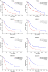
Diffuse gliomas are the most common type of primary brain and central nervous system (CNS) tumors. Protein disulfide isomerases (PDIs) such as P4HB and PDIA3 act as molecular chaperones for reconstructing misfolded proteins, and are involved in endoplasmic reticulum stress and the unfolded protein response. The present study focused on the role of P4HB and PDIA3 in diffuse gliomas. Analysis of GEO and HPA data revealed that the expression levels of P4HB and PDIA3 were upregulated in glioma datasets. their increased expression was then validated in 99 glioma specimens compared with 11 non-tumor tissues. High expression of P4HB and PDIA3 was significantly correlated with high Ki-67 and a high frequency of the TP53 mutation. Kaplan-Meier survival curve and Cox regression analyses showed that glioma patients with high P4HB and PDIA3 expression had a poor survival outcome, P4HB and PDIA3 could be independent prognostic biomarkers for diffuse gliomas. In vitro, knockdown of PDIA3 suppressed cell proliferation, induced cell apoptosis, and decreased the migration of glioma cells. Furthermore, downregulation of P4HB and PDIA3 may contribute to improve the survival of patients who receive chemotherapy and radiotherapy. The data suggest that high expression of P4HB and PDIA3 plays an important role in glioma progression, and could predict the survival outcome and therapeutic response of glioma patients. Therefore, protein disulfide isomerases may be explored as prognostic biomarkers and therapeutic targets for diffuse gliomas.
Figures


References
-
- Ostrom QT, Gittleman H, Fulop J, Liu M, Blanda R, Kromer C, Wolinsky Y, Kruchko C, Barnholtz-Sloan JS. CBTRUS Statistical Report: Primary Brain and Central Nervous System Tumors Diagnosed in the United States in 2008–2012. Neuro Oncol. 2015;17(Suppl 4):iv1–iv62. doi: 10.1093/neuonc/nov189. - DOI - PMC - PubMed
-
- Louis DN, Perry A, Reifenberger G, von Deimling A, Figarella-Branger D, Cavenee WK, Ohgaki H, Wiestler OD, Kleihues P, Ellison DW. The 2016 World Health Organization Classification of Tumors of the Central Nervous System: A summary. Acta Neuropathol. 2016;131:803–820. doi: 10.1007/s00401-016-1545-1. - DOI - PubMed
MeSH terms
Substances
LinkOut - more resources
Full Text Sources
Other Literature Sources
Medical
Research Materials
Miscellaneous

