Multiple Reaction Monitoring for the Quantitation of Serum Protein Glycosylation Profiles: Application to Ovarian Cancer
- PMID: 29207246
- PMCID: PMC6203861
- DOI: 10.1021/acs.jproteome.7b00541
Multiple Reaction Monitoring for the Quantitation of Serum Protein Glycosylation Profiles: Application to Ovarian Cancer
Abstract
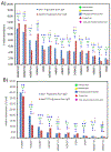
Protein glycosylation fingerprints are widely recognized as potential markers for disease states, and indeed differential glycosylation has been identified in multiple types of autoimmune diseases and several types of cancer. However, releasing the glycans leave the glycoproteins unknown; therefore, there exists a need for high-throughput methods that allow quantification of site- and protein-specific glycosylation patterns from complex biological mixtures. In this study, a targeted multiple reaction monitoring (MRM)-based method for the protein- and site-specific quantitation involving serum proteins immunoglobulins A, G and M, alpha-1-antitrypsin, transferrin, alpha-2-macroglobulin, haptoglobin, alpha-1-acid glycoprotein and complement C3 was developed. The method is based on tryptic digestion of serum glycoproteins, followed by immediate reverse phase UPLC-QQQ-MS analysis of glycopeptides. To quantitate protein glycosylation independent of the protein serum concentration, a nonglycosylated peptide was also monitored. Using this strategy, 178 glycopeptides and 18 peptides from serum glycoproteins are analyzed with good repeatability (interday CVs of 3.65-21-92%) in a single 17 min run. To assess the potential of the method, protein glycosylation was analyzed in serum samples from ovarian cancer patients and controls. A training set consisting of 40 cases and 40 controls was analyzed, and differential analyses were performed to identify aberrant glycopeptide levels. All findings were validated in an independent test set (n = 44 cases and n = 44 controls). In addition to the differential glycosylation on the immunoglobulins, which was reported previously, aberrant glycosylation was also observed on each of the glycoproteins, which could be corroborated in the test set. This report shows the development of a method for targeted protein- and site-specific glycosylation analysis and the potential of such methods in biomarker development.
Keywords: LC-MRM-MS; biomarker discovery; epithelial ovarian cancer; glycopeptides; protein glycosylation; protein quantitation; serum.
Conflict of interest statement
Conflict of Interest Statement
All authors have no financial or commercial conflicts of interest.
Figures


Similar articles
-
The Use of Multiple Reaction Monitoring on QQQ-MS for the Analysis of Protein- and Site-Specific Glycosylation Patterns in Serum.Methods Mol Biol. 2017;1503:63-82. doi: 10.1007/978-1-4939-6493-2_6. Methods Mol Biol. 2017. PMID: 27743359
-
A Method for Comprehensive Glycosite-Mapping and Direct Quantitation of Serum Glycoproteins.J Proteome Res. 2015 Dec 4;14(12):5179-92. doi: 10.1021/acs.jproteome.5b00756. Epub 2015 Nov 9. J Proteome Res. 2015. PMID: 26510530 Free PMC article.
-
Protein-Specific Differential Glycosylation of Immunoglobulins in Serum of Ovarian Cancer Patients.J Proteome Res. 2016 Mar 4;15(3):1002-10. doi: 10.1021/acs.jproteome.5b01071. Epub 2016 Feb 24. J Proteome Res. 2016. PMID: 26813784 Free PMC article.
-
Glycosylation changes on serum glycoproteins in ovarian cancer may contribute to disease pathogenesis.Dis Markers. 2008;25(4-5):219-32. doi: 10.1155/2008/601583. Dis Markers. 2008. PMID: 19126966 Free PMC article. Review.
-
Glycopeptide enrichment and separation for protein glycosylation analysis.J Sep Sci. 2012 Sep;35(18):2341-72. doi: 10.1002/jssc.201200434. J Sep Sci. 2012. PMID: 22997027 Review.
Cited by
-
A site-specific map of the human plasma glycome and its age and gender-associated alterations.Sci Rep. 2020 Oct 15;10(1):17505. doi: 10.1038/s41598-020-73588-x. Sci Rep. 2020. PMID: 33060657 Free PMC article.
-
N-Glycan and Glycopeptide Serum Biomarkers in Philippine Lung Cancer Patients Identified Using Liquid Chromatography-Tandem Mass Spectrometry.ACS Omega. 2022 Oct 26;7(44):40230-40240. doi: 10.1021/acsomega.2c05111. eCollection 2022 Nov 8. ACS Omega. 2022. PMID: 36385894 Free PMC article.
-
Quantitative Analysis of Sex-Hormone-Binding Globulin Glycosylation in Liver Diseases by Liquid Chromatography-Mass Spectrometry Parallel Reaction Monitoring.J Proteome Res. 2018 Aug 3;17(8):2755-2766. doi: 10.1021/acs.jproteome.8b00201. Epub 2018 Jul 16. J Proteome Res. 2018. PMID: 29972295 Free PMC article.
-
Distribution of de novo Donor-Specific Antibody Subclasses Quantified by Mass Spectrometry: High IgG3 Proportion Is Associated With Antibody-Mediated Rejection Occurrence and Severity.Front Immunol. 2020 Jun 2;11:919. doi: 10.3389/fimmu.2020.00919. eCollection 2020. Front Immunol. 2020. PMID: 32670261 Free PMC article.
-
Methods for quantification of glycopeptides by liquid separation and mass spectrometry.Mass Spectrom Rev. 2023 Mar;42(2):887-917. doi: 10.1002/mas.21771. Epub 2022 Feb 4. Mass Spectrom Rev. 2023. PMID: 35099083 Free PMC article. Review.
References
-
- Siegel R, Naishadham D, Jemal A, Cancer statistics, 2013. CA Cancer J Clin 2013, 63, 11–30. - PubMed
-
- Elschenbroich S, Ignatchenko V, Clarke B, Kalloger SE, et al., In-depth proteomics of ovarian cancer ascites: combining shotgun proteomics and selected reaction monitoring mass spectrometry. J Proteome Res 2011, 10, 2286–2299. - PubMed
-
- Woof JM, van Egmond M, Kerr MA, in: Mestecky J, Lamm ME, Strober W, Bienenstock J, et al. (Eds.), Mucosal Immunology, Elsevier, London: 2005, pp. 251–265.
Publication types
MeSH terms
Substances
Grants and funding
LinkOut - more resources
Full Text Sources
Other Literature Sources
Miscellaneous

