

Review
doi: 10.1002/art.40396.
Epub 2018 Feb 22.
Abnormal B Cell Development in Systemic Lupus Erythematosus: What the Genetics Tell Us
Affiliations
- PMID: 29207444
- PMCID: PMC5900717
- DOI: 10.1002/art.40396
Item in Clipboard
Review
Abnormal B Cell Development in Systemic Lupus Erythematosus: What the Genetics Tell Us
Arthritis Rheumatol.
2018 Apr.
No abstract available
Figures
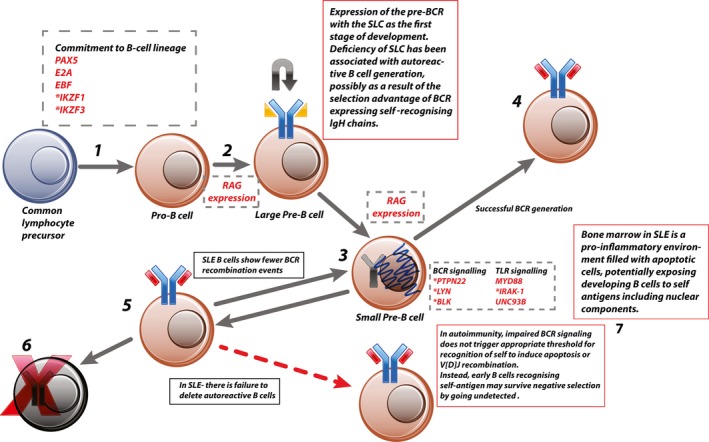
Central tolerance. 1, Common lymphocyte precursor commits to B cell lineage via expression of B cell–specific transcription factors (e.g., early B cell factor [EBF]), which initiates IgH rearrangement. 2, Expression of the generated IgH component of the pre–B cell receptor (pre‐BCR) is combined with the surrogate light chain (SLC). 3, Successful signaling through the pre‐BCR leads to a short burst of proliferation and internalization of the pre‐BCR and commences a second wave of recombination, this time in the light‐chain gene. 4, The generated BCR is then assessed for self‐recognition. Those cells that have generated non–self‐recognizing BCRs with functioning signaling switch off recombination‐activating gene (RAG) expression and become immature B cells. 5, Because V[D]J recombination is a stochastic process, a proportion of pre–B cells will generate autoreactive BCRs. This is detected by excess BCR signaling due to high‐affinity binding within the bone marrow or abundance of antigen. This leads to continued V[D]J recombination until acceptable BCR is generated or all possible recombination has been exhausted. 6, Failure to generate a non–self‐recognizing BCR leads to apoptosis. 7, In autoimmune disease this process is impaired, potentially by reduced signaling through the developing BCR, which fails to trigger the threshold for apoptosis. Genes or proteins involved at each stage are shown in dashed boxes. * = genes identified as risk‐associated loci in systemic lupus erythematosus (SLE). TLR = Toll‐like receptor; MyD88 = myeloid differentiation factor 88; IRAK‐1 = interleukin‐1 receptor–associated kinase 1; Unc‐93B = Unc‐93 homolog B.
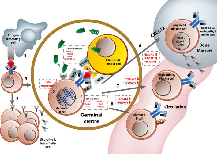
Peripheral tolerance. 1, Naive B cells in the marginal zone encounter their relevant antigen as presented by resident antigen‐presenting cells (APCs). 2, Some activated B cells remain outside the germinal center and become short‐lived low‐affinity antibody‐producing cells. 3, Activated B cells migrate to the germinal center (under influence of CXCL12 produced by bone marrow stromal cells), 4, where they interact with follicular helper T cells whose T cell receptors (TCRs) recognize self antigen. This also involves bidirectional signaling through multiple costimulatory molecules and the B cell receptor (BCR). 5, At this stage, most B cells undergo a round of somatic hypermutation to achieve affinity maturation. This requires expression of the RAG genes. Following this, activated B cells can differentiate into 3 potential cell types. 6, Long‐lived plasma cells are selected from the pool of B cells with the highest affinity receptors. They up‐regulate expression of CXCR4 and migrate toward their niche (usually in the bone marrow), where they reside and continue to produce background antibody. 7, Some activated cells terminally differentiate into high‐affinity plasma cells, which are responsible for the “second wave” of high‐affinity antibody after antigen exposure. 8, B cells with low‐affinity BCRs are preferentially selected to become memory B cells. Genes or proteins involved at each of the regulatory stages are shown in dashed boxes. * = genes identified as risk‐associated loci in systemic lupus erythematosus (SLE). IL‐21 = interleukin‐21; MHC = major histocompatibility complex; IL‐21R = IL‐21 receptor; RAG = recombination‐activating gene; ICOS = inducible costimulator; BANK‐1 = B cell scaffold protein with ankyrin repeats 1; UBE2L3 = ubiquitin‐conjugating enzyme E2 L3; BLIMP‐1 = B lymphocyte–induced maturation protein 1; XBP‐1 = X‐box binding protein 1.
References
-
- Kaul A, Gordon C, Crow MK, Touma Z, Urowitz MB, van Vollenhoven R, et al. Systemic lupus erythematosus. Nat Rev Dis Prim 2016;2:16039. - PubMed
-
- Deafen D, Escalante A, Weinrib L, Horwitz D, Bachman B, Roy‐Burman P, et al. A revised estimate of twin concordance in systemic lupus erythematosus. Arthritis Rheum 1992;35:311–8. - PubMed
-
- Iwata S, Tanaka Y. B‐cell subsets, signaling and their roles in secretion of autoantibodies. Lupus 2016;25:850–6. - PubMed
Publication types
MeSH terms
Grants and funding
LinkOut - more resources
Full Text Sources
Other Literature Sources
Medical
