

Isoxazole compound ML327 blocks MYC expression and tumor formation in neuroblastoma
- PMID: 29207623
- PMCID: PMC5710904
- DOI: 10.18632/oncotarget.19406
Isoxazole compound ML327 blocks MYC expression and tumor formation in neuroblastoma
Abstract
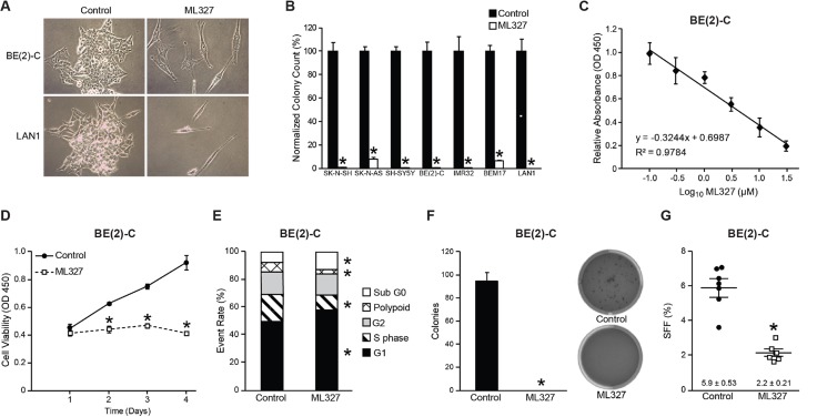
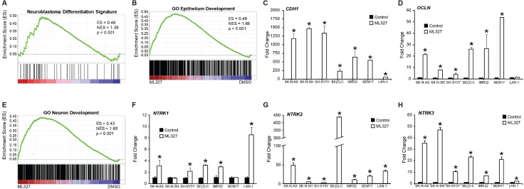
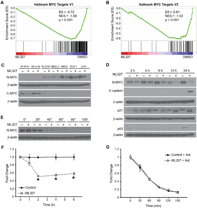
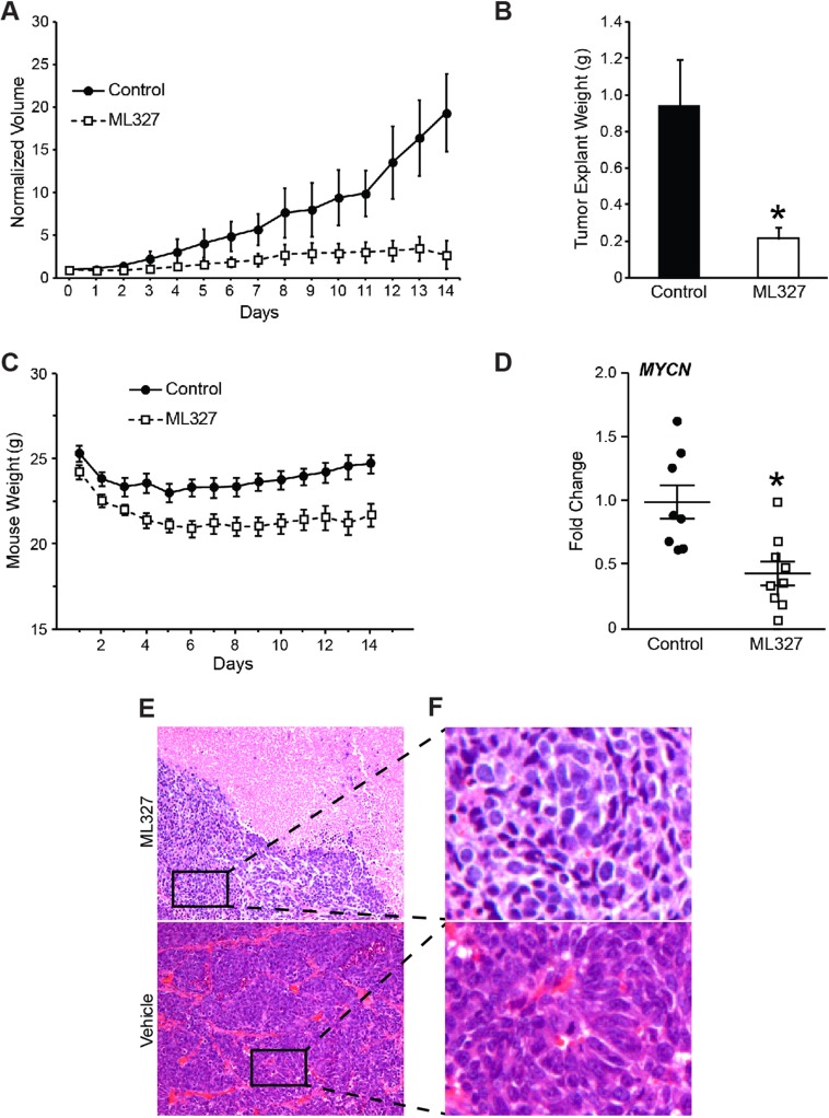
Neuroblastomas are the most common extracranial solid tumors in children and arise from the embryonic neural crest. MYCN-amplification is a feature of ∼30% of neuroblastoma tumors and portends a poor prognosis. Neural crest precursors undergo epithelial-to-mesenchymal transition (EMT) to gain migratory potential and populate the sympathoadrenal axis. Neuroblastomas are posited to arise due to a blockade of neural crest differentiation. We have recently reported effects of a novel MET inducing compound ML327 (N-(3-(2-hydroxynicotinamido) propyl)-5-phenylisoxazole-3-carboxamide) in colon cancer cells. Herein, we hypothesized that forced epithelial differentiation using ML327 would promote neuroblastoma differentiation. In this study, we demonstrate that ML327 in neuroblastoma cells induces a gene signature consistent with both epithelial and neuronal differentiation features with adaptation of an elongated phenotype. These features accompany induction of cell death and G1 cell cycle arrest with blockage of anchorage-independent growth and neurosphere formation. Furthermore, pretreatment with ML327 results in persistent defects in proliferative potential and tumor-initiating capacity, validating the pro-differentiating effects of our compound. Intriguingly, we have identified destabilization of MYC signaling as an early and consistent feature of ML327 treatment that is observed in both MYCN-amplified and MYCN-single copy neuroblastoma cell lines. Moreover, ML327 blocked MYCN mRNA levels and tumor progression in established MYCN-amplified xenografts. As such, ML327 may have potential efficacy, alone or in conjunction with existing therapeutic strategies against neuroblastoma. Future identification of the specific intracellular target of ML327 may inform future drug discovery efforts and enhance our understanding of MYC regulation.
Keywords: ML327; MYCN; epithelial-to-mesenchymal transition; neural crest; neuroblastoma.
Conflict of interest statement
CONFLICTS OF INTEREST The authors declare no potential conflicts of interest.
Figures

Similar articles
-
ML327 induces apoptosis and sensitizes Ewing sarcoma cells to TNF-related apoptosis-inducing ligand.Biochem Biophys Res Commun. 2017 Sep 16;491(2):463-468. doi: 10.1016/j.bbrc.2017.07.050. Epub 2017 Jul 14. Biochem Biophys Res Commun. 2017. PMID: 28716733 Free PMC article.
-
Distinct transcriptional MYCN/c-MYC activities are associated with spontaneous regression or malignant progression in neuroblastomas.Genome Biol. 2008 Oct 13;9(10):R150. doi: 10.1186/gb-2008-9-10-r150. Genome Biol. 2008. PMID: 18851746 Free PMC article.
-
Functional interplay between MYCN, NCYM, and OCT4 promotes aggressiveness of human neuroblastomas.Cancer Sci. 2015 Jul;106(7):840-7. doi: 10.1111/cas.12677. Epub 2015 May 19. Cancer Sci. 2015. PMID: 25880909 Free PMC article.
-
The MYCN oncoprotein as a drug development target.Cancer Lett. 2003 Jul 18;197(1-2):125-30. doi: 10.1016/s0304-3835(03)00096-x. Cancer Lett. 2003. PMID: 12880971 Review.
-
The MYCN oncogene and differentiation in neuroblastoma.Semin Cancer Biol. 2011 Oct;21(4):256-66. doi: 10.1016/j.semcancer.2011.08.001. Epub 2011 Aug 9. Semin Cancer Biol. 2011. PMID: 21849159 Review.
Cited by
-
A Novel Pharmacological Approach to Enhance the Integrity and Accelerate Restitution of the Intestinal Epithelial Barrier.Inflamm Bowel Dis. 2020 Aug 20;26(9):1340-1352. doi: 10.1093/ibd/izaa063. Inflamm Bowel Dis. 2020. PMID: 32266946 Free PMC article.
-
GDP-mannose 4,6-dehydratase is a key driver of MYCN-amplified neuroblastoma core fucosylation and tumorigenesis.Oncogene. 2025 May;44(18):1272-1283. doi: 10.1038/s41388-025-03297-0. Epub 2025 Feb 16. Oncogene. 2025. PMID: 39956863 Free PMC article.
-
ML327 induces apoptosis and sensitizes Ewing sarcoma cells to TNF-related apoptosis-inducing ligand.Biochem Biophys Res Commun. 2017 Sep 16;491(2):463-468. doi: 10.1016/j.bbrc.2017.07.050. Epub 2017 Jul 14. Biochem Biophys Res Commun. 2017. PMID: 28716733 Free PMC article.
References
-
- Park JR, Bagatell R, London WB, Maris JM, Cohn SL, Mattay KK, Hogarty M. Children’s Oncology Group’s 2013 blueprint for research: neuroblastoma. Pediatr Blood Cancer. 2013;60:985–993. - PubMed
-
- Brodeur GM. Neuroblastoma: biological insights into a clinical enigma. Nat Rev Cancer. 2003;3:203–216. - PubMed
Grants and funding
LinkOut - more resources
Full Text Sources
Other Literature Sources
Molecular Biology Databases
Research Materials
Miscellaneous
