. 2017 Aug 24;8(53):91209-91222.
doi: 10.18632/oncotarget.20436. eCollection 2017 Oct 31.

Wheat germ agglutinin-induced paraptosis-like cell death and protective autophagy is mediated by autophagy-linked FYVE inhibition

Affiliations

Wheat germ agglutinin-induced paraptosis-like cell death and protective autophagy is mediated by autophagy-linked FYVE inhibition

Tsung Lin Tsai et al. Oncotarget. .

Abstract

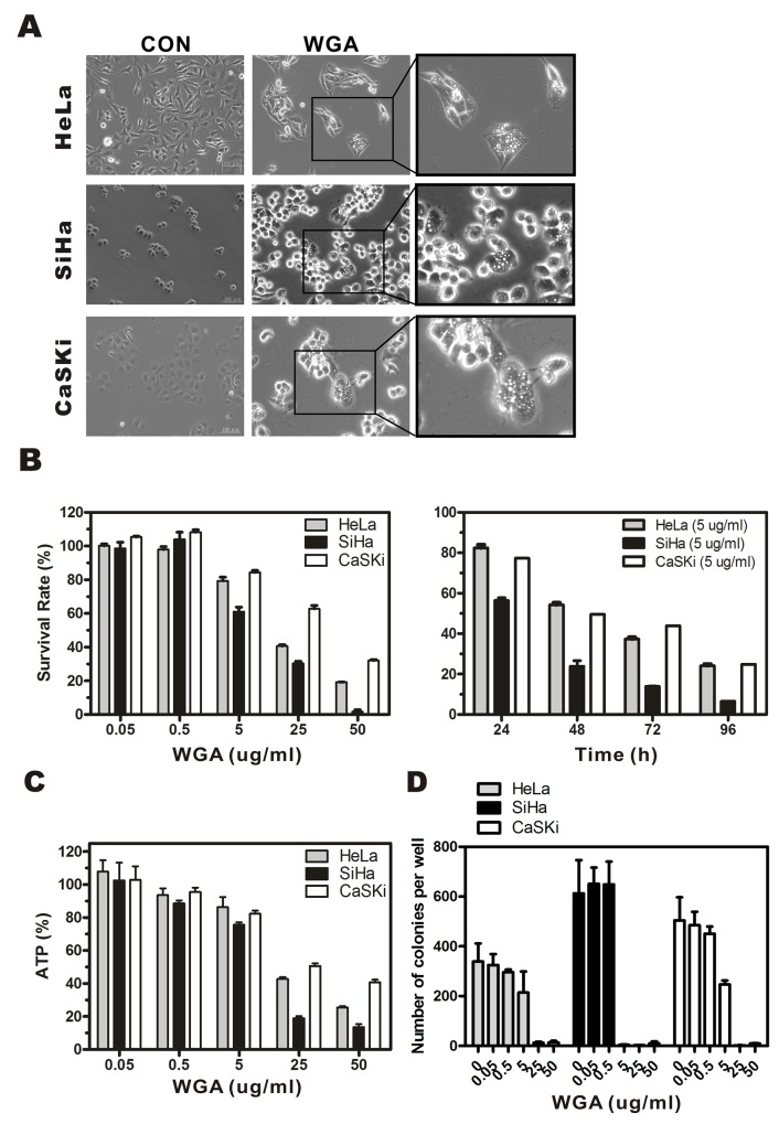
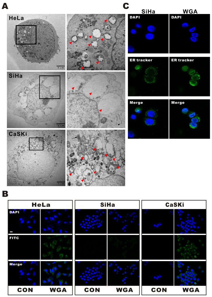
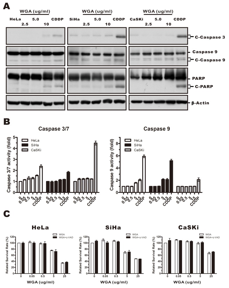
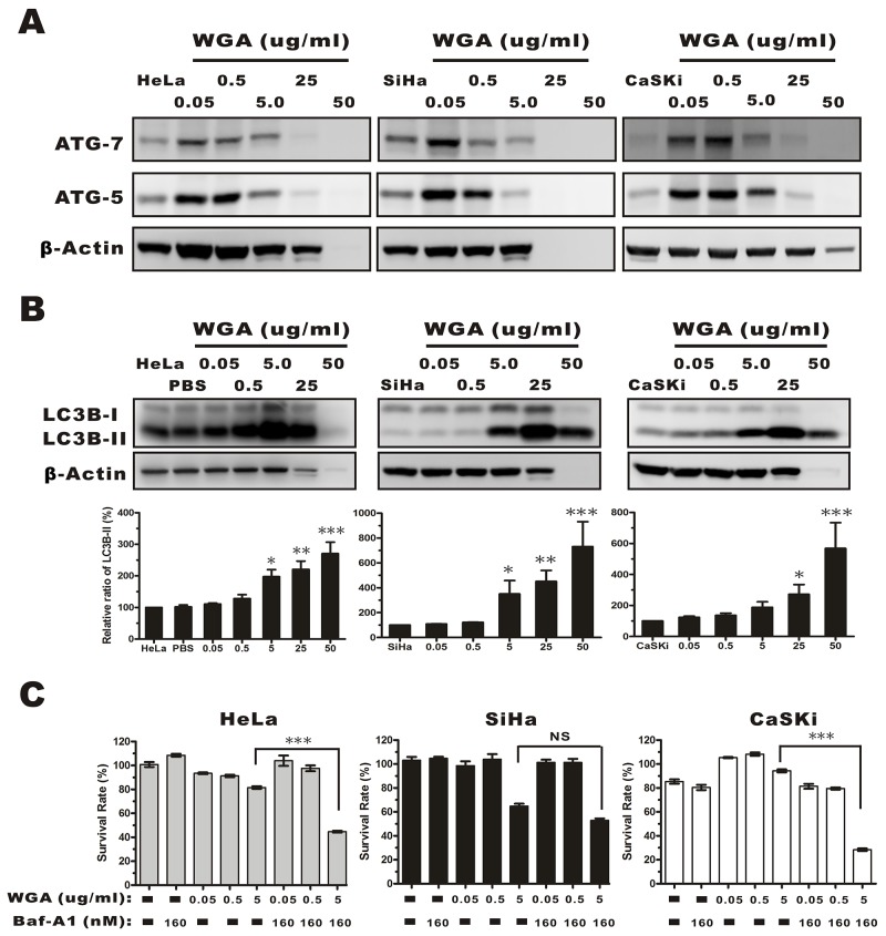
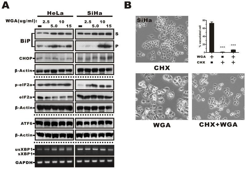
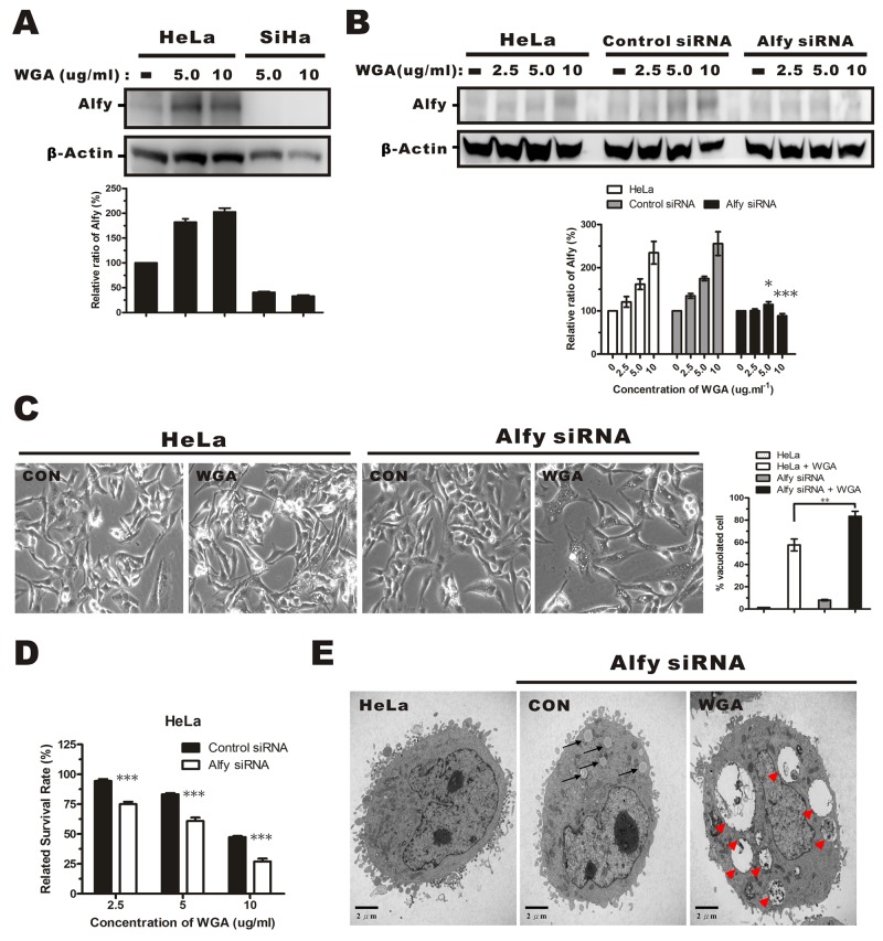
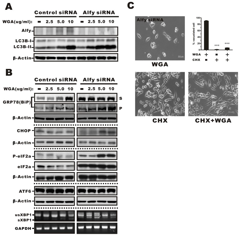
Wheat germ agglutinin (WGA) is a lectin that specifically binds cell surface glycoproteins and disrupts nuclear pore complex function through its interaction with POM121. Our data indicate WGA induces paraptosis-like cell death without caspase activation. We observed the main features of paraptosis, including cytoplasmic vacuolation, endoplasmic reticulum dilation and increased ER stress, and the unfolded protein response in WGA-treated cervical carcinoma cells. Conversion of microtubule-associated protein I light chain 3 (LC3-I) into LC3-II and punctuate formation suggestive of autophagy were observed in WGA-treated cells. WGA-induced autophagy antagonized paraptosis in HeLa and CaSKi cells, which expressed autophagy-linked FYVE (Alfy) protein, but not in SiHa cells that did not express Alfy. Alfy knockdown in HeLa cells induced paraptosis-like cell death. These data indicate that WGA-induced cell death occurs through paraptosis and that autophagy may exert a protective effect. WGA treatment and Alfy inhibition could be an effective therapeutic strategy for apoptosis-resistant cervical cancer cells.

Keywords: Alfy; cell death; cytoplasmic vacuolation; paraptosis; wheat germ agglutinin.


Conflict of interest statement

CONFLICTS OF INTEREST The authors declare that there are no conflicts of interest.

Figures

Figure 1
Figure 1. WGA induces formation of cytoplasmic vacuoles and paraptosis-like cell death in cervical carcinoma cells
(A) Light micrographs of WGA-treated and untreated control cancer cells. HeLa cells, SiHa cells, and CaSKi cells were treated with WGA (10, 5, and 20 μg/mL, respectively). (B) MTT assays of cell viability. Cells were incubated with WGA at different concentrations for 24 h, or with a fixed concentration of 5 μg/mL for different lengths of time. (C) ATP levels 24 h after treatment with WGA at the indicated concentrations. (D) Clonogenic assays showing decreased viability of HeLa, SiHa, and CaSKi cells after treatment with WGA at concentrations ranging from 0.05–50 μg/mL. After long-term incubation (10–14 days), cells were fixed and stained with crystal violet, and the number of colonies counted. Data are expressed as the mean ± SD based on three independent experiments.
Figure 2
Figure 2. WGA induces both paraptosis and autophagy in HeLa and CaSKi cells, but only paraptosis in SiHa cells
(A) TEM images of HeLa, SiHa, and CaSKi cells treated with WGA for 24 h. The inset shows a part of the image at higher magnification. The red arrow heads point to extensive, clear paraptosis vacuoles. The black arrows point to autophagosomes or autolysosomes. (B) Autophagic vacuoles are stained with Cyto-ID® green fluorescent dyeandthenuclei with DAPI. An increase in green fluorescence was observed in WGA-treated HeLa and CaSKi cells. Little to no staining of vacuoles was observed in SiHa or control cells. (C) Confocal images of SiHa cells with and without WGA treatment for 24 h. The ER structures are stained with ER-tracker and the nuclei with DAPI.
Figure 3
Figure 3. WGA-induced cytoplasmic vacuolation and cell death was not associated with caspase-dependent apoptosis
(A) HeLa, SiHa, and CaSKi cells were treated with 2.5, 5, or 10 μg/mL WGA for 24 h, and the expression of caspase-3, -9, and PARP measured by western blot. β-Actin was used as a loading control. (B) The activity of caspase-3/7 and caspase-9 was detected using Caspase-Glo assays. Data are expressed as the mean ± SD for four replicates. (C) HeLa, SiHa, and CaSKi cells (untreated or pre-treated with 25 μM z-VAD-fmk) were treated with WGA at the indicated concentrations. Cell survival was assessed using MTT assays. Data are expressed the mean ± SD for six duplicated experiments.
Figure 4
Figure 4. Evaluation of autophagy by monitoring ATG-5, -7, and the conversion LC3B-I to LC3B-II in whole protein extracts from HeLa, SiHa, and CaSKi cells treated with WGA at the indicated concentrations
(A) ATG-5 and -7 expression increased in all cervical carcinoma cells following treatment with WGA at the indicated concentrations. (B) Western blot showing dose-dependent expression of LC3B in HeLa, SiHa, and CaSKi cells treated with WGA for 24 h. The cytoplasmic form of LC3 (LC3B-I) and the autophagosomal membrane-bound form (LC3B-II) were both detected. We quantified the relative levels of LC3-II to β-Actin. Bars represent the mean ± SD of three independent experiments (*, P < 0.05; **, P < 0.01; ***, P < 0.001). (C) MTT assays in HeLa, SiHa, and CaSKi cells treated with the indicated concentrations of WGA in the presence and absence of Baf-A1, an inhibitor of autophagy. Bars represent the mean ± SD of four independent experiments (P > 0.05, not significant; ***, P < 0.001).
Figure 5
Figure 5. ER stress-mediated activation of the UPR in WGA-treated cervical carcinoma cells and prevention of vacuolation by CHX
(A) Western blot analysis showing increased expression of markers of ER stress, including phosphorylated eIF2α, BiP, and ER chaperones in the Triton-soluble (S) or -insoluble pellet fractions (P), and the transcription factor CHOP in WGA-treated cancer cells. No changes in ATF6 expression or XBP1 splicing were observed in response to WGA treatment. (B) Inhibition of cytoplasmic vacuolation after the addition of CHX. SiHa cells were treated with either WGA (5 μg/mL) alone or in combination with CHX (25 μM) for 1.5 h and the percentage of vacuolated cells determined by counting at least 200 cells in three independent experiments. Bars represent the mean ± SD (***, P < 0.001).
Figure 6
Figure 6. Effects of Alfy knockdown on WGA-induced cytoplasmic vacuolation and cell death
(A) Western blotsof total cell lysates showing increased Alfy expression in HeLa cells, but not SiHa cells, after treatment with WGA (5.0 and 10 μg/mL) for 24 h. (B) Western blot showing Alfy expression in WGA-treated HeLa cells following knockdown of Alfy with shRNA. Empty vector (control siRNA) was used as negative control. The fold change in Alfy expression after WGA treatment was quantified using NIH ImageJ. The relative units were normalized to β-Actin and compared to untreated control cells. Data are expressed as the mean ± SD for three independent experiments. (*, P < 0.05; ***, P < 0.001) (C) Phase-contrast images showing the effects of Alfy knockdown on WGA-induced cytoplasmic vacuolation. Bar graph showing the percentage of vacuolated HeLa cells expressing control siRNA or Alfy siRNA with or without WGA treatment. At least 200 cells were counted in three independent experiments. Bars represent the mean ± SD (**, P < 0.01). (D) MTT assays of cell viability after treatment of Alfy knockdown and control HeLa cells with or without WGA for 24 h. Data represent an average of three independent experiments. Bars represent the mean ± SD (***, P < 0.001). (E) Formation of cytoplasmic vacuoles in HeLa cells following Alfy knockdown. Cells were transfected with Alfy siRNA and treated for 24 h with or without WGA (10 μg/mL). The black arrow head points to small vacuoles and the red arrow head points to extensive paraptosis-like vacuoles.
Figure 7
Figure 7. Effects of WGA on Alfy expression and the effects of Alfy knockdown ER stress-mediated cytoplasmic vacuolation and the UPR
(A) Expression of Alfy andLC3 in HeLa cells in Alfy knockdown or vector control cells measured by Western blotting following treatment with the indicated concentrations of WGA. β-Actin was used as a loading control. (B) Markers of ER stress were evaluated in Alfy knockdown and vector control HeLa cells. Levels of phosphorylated eIF2α, BiP, ER chaperones, and CHOP in the Triton-soluble fraction (S) or -insoluble pellet (P) fraction in Alfy knockdown HeLa cells treated with WGA. No change in ATF6 expression or XBP1 splicing were observed in response to WGA treatment in Alfy knockdown or control HeLa cells. (C) Inhibition of cytoplasmic vacuolation in response to CHX. Alfy knockdown HeLa cells were treated with WGA (5 μg/mL) alone or in combination with CHX (25 μM) for 1.5 h and the percentage vacuolated cells calculated by counting at least 200 cells in three independent experiments. Bars represent the mean ± SD (***, P < 0.001).

Similar articles

Cited by

  • Glycosylation-Dependent Induction of Programmed Cell Death in Murine Adenocarcinoma Cells.
    Parshenkov A, Hennet T. Parshenkov A, et al. Front Immunol. 2022 Feb 10;13:797759. doi: 10.3389/fimmu.2022.797759. eCollection 2022. Front Immunol. 2022. PMID: 35222379 Free PMC article.
  • Paraptosis: a unique cell death mode for targeting cancer.
    Hanson S, Dharan A, P V J, Pal S, Nair BG, Kar R, Mishra N. Hanson S, et al. Front Pharmacol. 2023 Jun 15;14:1159409. doi: 10.3389/fphar.2023.1159409. eCollection 2023. Front Pharmacol. 2023. PMID: 37397502 Free PMC article. Review.
  • Guidelines for the use and interpretation of assays for monitoring autophagy (4th edition)1.
    Klionsky DJ, Abdel-Aziz AK, Abdelfatah S, Abdellatif M, Abdoli A, Abel S, Abeliovich H, Abildgaard MH, Abudu YP, Acevedo-Arozena A, Adamopoulos IE, Adeli K, Adolph TE, Adornetto A, Aflaki E, Agam G, Agarwal A, Aggarwal BB, Agnello M, Agostinis P, Agrewala JN, Agrotis A, Aguilar PV, Ahmad ST, Ahmed ZM, Ahumada-Castro U, Aits S, Aizawa S, Akkoc Y, Akoumianaki T, Akpinar HA, Al-Abd AM, Al-Akra L, Al-Gharaibeh A, Alaoui-Jamali MA, Alberti S, Alcocer-Gómez E, Alessandri C, Ali M, Alim Al-Bari MA, Aliwaini S, Alizadeh J, Almacellas E, Almasan A, Alonso A, Alonso GD, Altan-Bonnet N, Altieri DC, Álvarez ÉMC, Alves S, Alves da Costa C, Alzaharna MM, Amadio M, Amantini C, Amaral C, Ambrosio S, Amer AO, Ammanathan V, An Z, Andersen SU, Andrabi SA, Andrade-Silva M, Andres AM, Angelini S, Ann D, Anozie UC, Ansari MY, Antas P, Antebi A, Antón Z, Anwar T, Apetoh L, Apostolova N, Araki T, Araki Y, Arasaki K, Araújo WL, Araya J, Arden C, Arévalo MA, Arguelles S, Arias E, Arikkath J, Arimoto H, Ariosa AR, Armstrong-James D, Arnauné-Pelloquin L, Aroca A, Arroyo DS, Arsov I, Artero R, Asaro DML, Aschner M, Ashrafizadeh M, Ashur-Fabian O, Atanasov AG, Au AK, Auberger P, Auner HW, Aurelian L, Autelli R… See abstract for full author list ➔ Klionsky DJ, et al. Autophagy. 2021 Jan;17(1):1-382. doi: 10.1080/15548627.2020.1797280. Epub 2021 Feb 8. Autophagy. 2021. PMID: 33634751 Free PMC article.
  • Nuclear pore protein POM121 regulates subcellular localization and transcriptional activity of PPARγ.
    Yu Y, Farooq MS, Eberhart Meessen S, Jiang Y, Kato D, Zhan T, Weiss C, Seger R, Kang W, Zhang X, Yu J, Ebert MPA, Burgermeister E. Yu Y, et al. Cell Death Dis. 2024 Jan 4;15(1):7. doi: 10.1038/s41419-023-06371-1. Cell Death Dis. 2024. PMID: 38177114 Free PMC article.
  • Wheat Germ Agglutinin as a Potential Therapeutic Agent for Leukemia.
    Ryva B, Zhang K, Asthana A, Wong D, Vicioso Y, Parameswaran R. Ryva B, et al. Front Oncol. 2019 Feb 21;9:100. doi: 10.3389/fonc.2019.00100. eCollection 2019. Front Oncol. 2019. PMID: 30847305 Free PMC article.

References

    1. Hallberg E, Wozniak RW, Blobel G. An integral membrane protein of the pore membrane domain of the nuclear envelope contains a nucleoporin-like region. J Cell Biol. 1993;122:513–521. - PMC - PubMed
    1. Pante N, Jarmolowski A, Izaurralde E, Sauder U, Baschong W, Mattai IW. Visualizing nuclear export of different classes of RNA by electron microscopy. RNA. 1997;3:498–513. - PMC - PubMed
    1. Meyer T, Begitt A, Lodige I, Rossum MV, Vinkemeier U. Constitutive and IFN-γ-induced nuclear import of STAT1 proceed through independent pathway. EMBO J. 2002;21:344–354. - PMC - PubMed
    1. Li XT, Ju RJ, Li XY, Zeng F, Shi JF, Liu L, Zhang CX, Sun MG, Lou JN, Lu WL. Multifunctional targeting daunorubicin plus quinacrine liposomes, modified by wheat germ agglutinin and tamoxifen, for treating brain glioma and glioma stem cells. Oncotarget. 2014;5:6497–6511. doi: 10.18632/oncotarget.2267. - DOI - PMC - PubMed
    1. Meng Y, Hou X, Lei J, Chen M, Cong S, Zhang Y, King W, Li G, Li X. Multi-function liposomes enhancing target and antibacterial immunity for antimicrobial and anti-biofilm against methicillin-resistant staphylococcus aureus. Pharm Res. 2016;33:763–75. - PubMed

LinkOut - more resources