. 2017 Aug 24;8(53):91306-91315.
doi: 10.18632/oncotarget.20444. eCollection 2017 Oct 31.

Nitidine chloride acts as an apoptosis inducer in human oral cancer cells and a nude mouse xenograft model via inhibition of STAT3

Affiliations

Nitidine chloride acts as an apoptosis inducer in human oral cancer cells and a nude mouse xenograft model via inhibition of STAT3

Lee-Han Kim et al. Oncotarget. .

Abstract

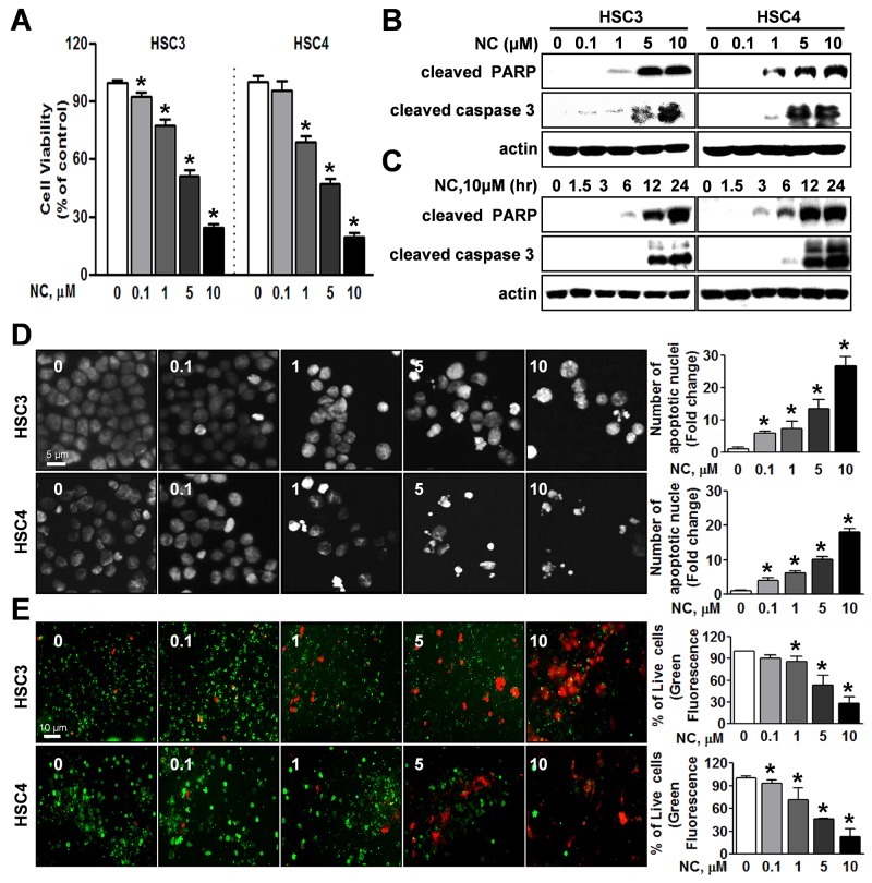
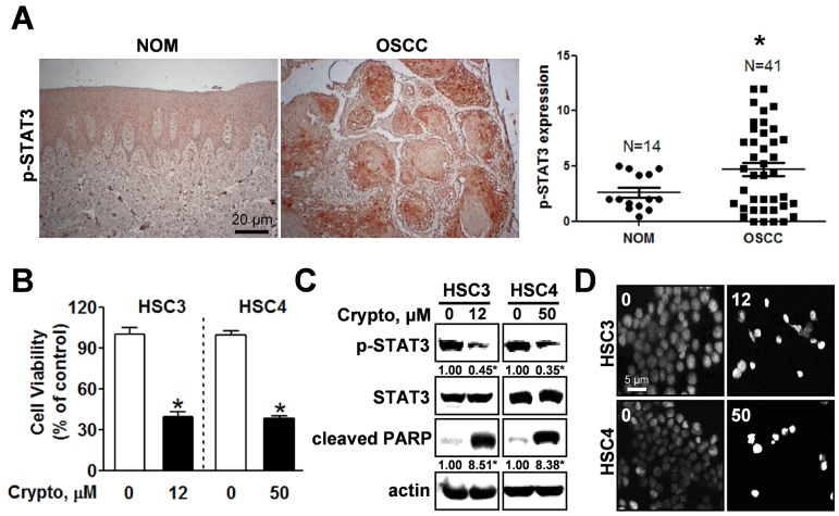
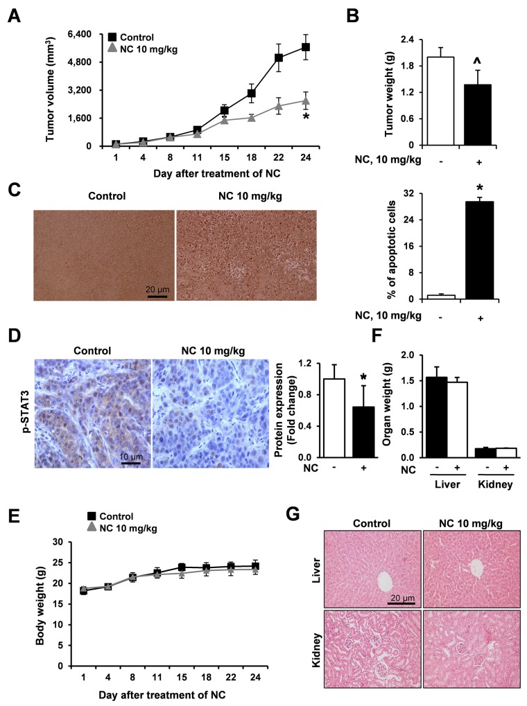
Nitidine chloride (NC) is a natural alkaloid compound derived from the plant Zanthoxylum nitidum and is known for its therapeutic anticancer potential. In this study, we investigated the effects of NC on growth and signaling pathways in human oral cancer cell lines and a tumor xenograft model. The apoptotic effects and related molecular targets of NC on human oral cancer were investigated using trypan blue exclusion assay, DAPI staining, Live/Dead assay, Western blotting, Immunohistochemistry/Immunofluorescence and a nude mouse tumor xenograft. NC decreased cell viability in both HSC3 and HSC4 cell lines; further analysis demonstrated that cell viability was reduced via apoptosis. STAT3 was hyper-phosphorylated in human oral squamous cell carcinoma (OSCC) compared with normal oral mucosa (NOM) and dephosphorylation of STAT3 by the potent STAT3 inhibitor, cryptotanshinone or NC decreased cell viability and induced apoptosis. NC also suppressed cell viability and induced apoptosis accompanied by dephosphorylating STAT3 in four other oral cancer cell lines. In a tumor xenograft model bearing HSC3 cell tumors, NC suppressed tumor growth and induced apoptosis by regulating STAT3 signaling without liver or kidney toxicity. Our findings suggest that NC is a promising chemotherapeutic candidate against human oral cancer.

Keywords: STAT3; apoptosis; nitidine chloride; oral cancer.


Conflict of interest statement

CONFLICTS OF INTEREST No potential conflicts of interest are declared.

Figures

Figure 1
Figure 1. Effect of nitidine chloride (NC) on cell viability and apoptosis in human oral cancer cell lines
HSC3 and HSC4 cells were treated with DMSO or various concentrations of NC (0.1, 1, 5 and 10 μM) for 24 hr or with 10 μM of NC for 1.5, 3, 6, 12 and 24 hr. (A) The effect of NC on cell viability was determined using a trypan blue exclusion assay. The graphs are expressed the mean ± S.D. of triplicate experiments and significance (p < 0.05) compared with the DMSO-treated group was indicated (*). (B and C) The apoptotic effect of NC was determined by Western blotting with the indicated antibodies (cleaved PARP and caspase 3). Actin was used as an internal control. (D) Fluorescence microscopy images of 4’-6-diamidino-2-phenylindole (DAPI)-stained HSC3 and HSC4 cells (magnification, X400). The number of cells with nuclear condensation and fragmentation was quantified. The graphs represent the mean ± S.D. of triplicate experiments. *, p < 0.05 is compared with control group. (E) Qualitative assessments of NC-induced cell death by a live/dead assay, which differentially labels live (green) and dead (red) cells with fluorescent dyes (magnification, X200). The graphs represent the mean ± S.D. of triplicate experiments. *, p < 0.05 is compared with control group.
Figure 2
Figure 2. STAT3 is hyper-phosphorylated in OSCC and inhibiting phosphorylation of STAT3 by cryptotanshione triggers apoptosis in oral cancer cell lines
(A) Left panel: Expression of phosphorylated STAT3 (p-STAT3) was evaluated by immunohistochemistry in tissue samples of patients with OSCC (n=41) compared with normal oral mucosa (NOM, n=14); Right panel: Dot-plot graph of p-STAT3 expression. (*) indicates p < 0.05 significant difference between NOM and OSCC group. (B) HSC3 and HSC4 cells were treated with DMSO or the STAT3 inhibitor, cryptotanshinone (Crypto, 12 and 50 μM, respectively) and cell viability was analyzed using a trypan blue exclusion assay. The graphs represent the mean ± S.D. of triplicate experiments. *, p < 0.05 is compared with control group. (C) Whole-cell lysates were analyzed by Western blotting using antibodies against p-STAT3, STAT3, and cleaved PARP. Data represent the mean of triplicate experiments. *, p < 0.05 is compared with control group. (D) Nuclear condensation and fragmentation were assessed by staining with DAPI (magnification X400).
Figure 3
Figure 3. Nitidine chloride (NC) reduces the expression levels of p-STAT3 in oral cancer cell lines
The expression of p-STAT3 was determined by Western blotting in a concentration- (A) and time-dependent manner (B). Data represent the mean of triplicate experiments. *, p < 0.05 is compared with control group. (C) p-STAT3 protein expression was confirmed by immunofluorescence using florescence microscopy (magnification X200).
Figure 4
Figure 4. The effect of NC on cell growth and apoptosis in four other different oral cancer cell lines (YD15, MC3, HN22 and Ca9.22)
Cells were treated with various concentrations of NC for 24hr. The growth inhibitory effect of NC was examined by trypan blue exclusion assay, and the effects of NC on PARP and caspase 3 cleavages, STAT3, and p-STAT3 were analyzed by Western blotting. The graphs represent the mean ± S.D. of triplicate experiments. *, p < 0.05 are compared with control group.
Figure 5
Figure 5. NC suppresses tumor growth and induces apoptosis in a nude mouse xenograft model bearing HSC3 cells
Athymic nude mice bearing HSC3 cells (4 mice per group) were treated with vehicle control or NC (10 mg/kg) for 24 days. Tumor volume (A), tumor weight (B) and body weight (E) were calculated at the indicated time points after treatment with NC. The graphs represent the mean ± S.E. of triplicate experiments. *, p < 0.05 is compared with control group. ^p=0.057 (C) Apoptosis was detected in tumor tissues using TUNEL assay (magnification, X200). Results are expressed as mean ± S.E and significance (p < 0.05) compared with the control-treated controls are indicated (*). (D) The expression levels of p-STAT3 were evaluated using immunohistochemistry. Slides were observed using a microscope (magnification X400). The graphs represent the mean ± S.E. of triplicate experiments. *, p < 0.05 is compared with control group. (F) Organ weights (liver and kidney) were measured. (G) Two organ tissue samples from control- and NC-treated mice were subjected to histopathological analysis using H&E staining (magnification, X200).

Similar articles

Cited by

References

    1. Fang Z, Tang Y, Jiao W, Xing Z, Guo Z, Wang W, Shi B, Xu Z, Liu Z. Nitidine chloride inhibits renal cancer cell metastasis via suppressing AKT signaling pathway. Food Chem Toxicol. 2013;60:246–251. - PubMed
    1. Wang Z, Jiang W, Zhang Z, Qian M, Du B. Nitidine chloride inhibits LPS-induced inflammatory cytokines production via MAPK and NF-kappaB pathway in RAW 264.7 cells. J Ethnopharmacol. 2012;144:145–150. - PubMed
    1. Chen J, Wang J, Lin L, He L, Wu Y, Zhang L, Yi Z, Chen Y, Pang X, Liu M. Inhibition of STAT3 signaling pathway by nitidine chloride suppressed the angiogenesis and growth of human gastric cancer. Mol Cancer Ther. 2012;11:277–287. - PubMed
    1. Kang M, Ou H, Wang R, Liu W, Tang A. The effect of nitidine chloride on the proliferation and apoptosis of nasopharyngeal carcinoma cells. J BUON. 2014;19:130–136. - PubMed
    1. Sun M, Zhang N, Wang X, Cai C, Cun J, Li Y, Lv S, Yang Q. Nitidine chloride induces apoptosis, cell cycle arrest, and synergistic cytotoxicity with doxorubicin in breast cancer cells. Tumour Biol. 2014;35:10201–10212. - PubMed

LinkOut - more resources