. 2017 Dec 6;17(1):827.
doi: 10.1186/s12885-017-3823-2.

Decreased expression of the β2 integrin on tumor cells is associated with a reduction in liver metastasis of colorectal cancer in mice

Affiliations

Decreased expression of the β2 integrin on tumor cells is associated with a reduction in liver metastasis of colorectal cancer in mice

Aitor Benedicto et al. BMC Cancer. .

Abstract

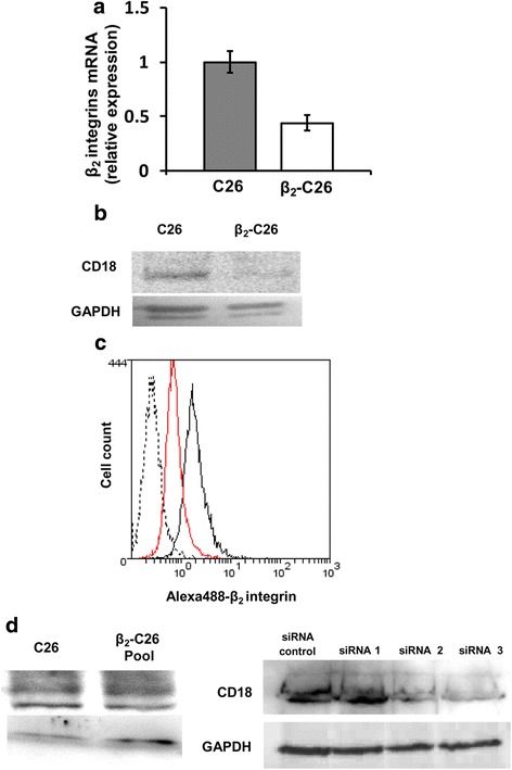
Background: Lymphocyte Function-Associated Antigen-1 (LFA-1; CD18/CD11a) is one of the main adhesion molecules used by immune cells to infiltrate the liver under inflammatory conditions. Recently, the expression of this integrin has also been reported on several solid tumors, including colorectal cancer. However, its functional role in the metastatic progression to the liver remains unknown. Using in vitro assays and an experimental orthotopic in vivo model of liver metastasis, we aimed to elucidate the role of tumor LFA-1 in the metastatic progression by means of the partial depletion of the β2 subunit of LFA-1, required for integrin activation, firm adhesion and signaling.

Methods: To do so, we evaluated the effects of β2 reduction on the murine colon carcinoma C26 cell line on their pro-metastatic features in vitro and their metastatic potential in vivo in a mouse model of colon carcinoma metastasis to the liver.

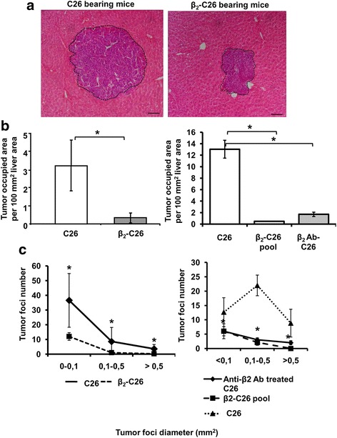
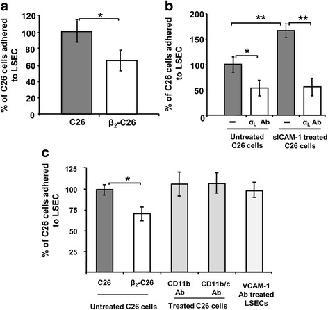
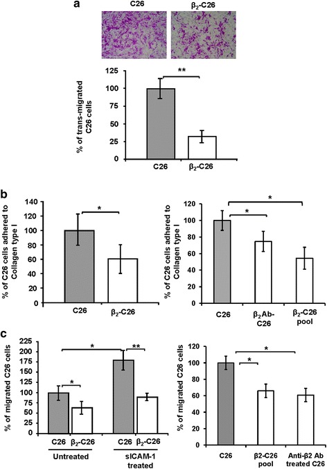
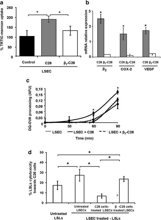
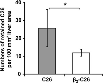
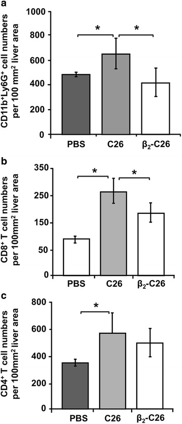
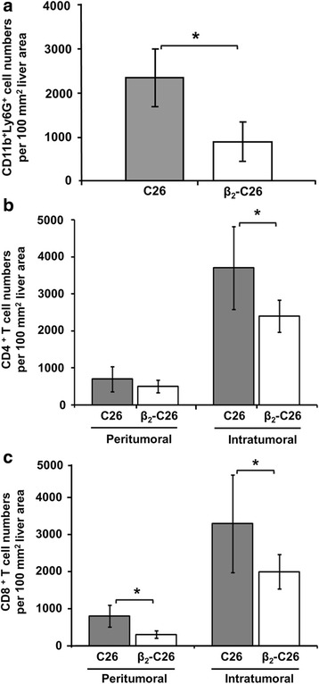
Results: The reduction in β2 integrin expression correlated with a slower proliferation, and a reduced adhesion and migration of C26 cells in an in vitro setting. Additionally, tumor cells with a reduced in β2 integrin expression were unable to activate the liver sinusoidal endothelial cells (LSECs). This resulted in a recovery of the cytotoxic potential of liver lymphocytes which is compromised by LSECs activated by C26 cells. This was related to the abrogation of RNA expression of inflammatory and angiogenic cytokines by C26 cells after their activation with sICAM-1, the main ligand of β2αL. Furthermore, in vivo tumor cell retention and metastasis were profoundly reduced, along with a decrease in the recruitment and infiltration of myeloid derived suppressor cells (MDSCs) and lymphocytes to the liver.

Conclusion: Taken together, our findings uncovered the modulatory role for the tumor β2 subunit of the LFA-1 integrin in the metastatic progression of colorectal cancer to the liver by impairing activation of liver endothelium and thus, the local immune response in the liver. Besides, this integrin also showed to be critical in vivo for tumor cell retention, cytokine release, leukocyte recruitment and metastasis development. These data support a therapeutical potential of the integrin LFA-1 as a target for the treatment of colorectal liver metastasis.

Keywords: Colorectal cancer; Endothelial cells; ICAM-1; Immune response; LFA-1; Liver metastasis; Tumor microenvironment; β2 integrin.


Conflict of interest statement

Ethics approval

All the animal proceedings were approved by the Basque Country University Ethical Committee (CEID) in accordance with institutional, national and international guidelines regarding the protection and care of animals use for scientific purposes.

Consent for publication

No applicable.

Competing interests

The authors declare that they have no competing interests.

Publisher’s Note

Springer Nature remains neutral with regard to jurisdictional claims in published maps and institutional affiliations.

Figures

Fig. 1
Fig. 1
Analysis of the expression of β2-integrin on C26 and β2-C26 cells. Lysates of unmodified (wild type) and β2-depleted C26 cells were collected for RNA (a) and protein (b-c) expression levels. a For β2 integrin RNA analysis by RT-PCR the following primers were used: forward ATCCTGACCCCCAATGATGG, reverse 5’CGGATGGGTAGTCGAACTCA. GAPDH was used as an internal control (house keeping gene) (b) Protein lysates were obtained from 1 clone cell line clones as described in Material and methods. At the protein level, the β2 integrin was detected by Western Blotting applying specific antibodies recognizing the β2 subunit of the integrin. c Tumor cells were incubated in the presence of specific antibodies for the integrin β2. Then, cells were subjected to FACS analysis after incubation of Alexa-488 antibody. The black line represents C26 cells, the red line represents β2-C26 cells and dash line represents the negative control. d Protein lysates were obtained from a pool of stably transfected C26 (left). Additionally, protein lysates were obtained from C26 tumor cells transiently transfected either with a control siRNA or three siRNAs specific for β2 integrin (right). At the protein level, the β2 integrin was detected by Western Blotting applying specific antibodies recognizing the β2 subunit of the integrin
Fig. 2
Fig. 2
Effect of the partial deficiency of the integrin subunit β2 on C26 colon carcinoma cells on tumor metastasis to the liver. Mice were sacrificed 14 days after either C26, β2-C26 cells i.s. inoculation, and metastatic development was quantified in paraffin embedded liver sections. In some experiments the mice were inoculated with a pool of β2-C26 cells or with C26 pre-treated with a neutralizing antibody for β2 integrin. a H&E staining showing a reduced size of metastatic foci in animals injected with β2-C26 cells versus C26 cells. b The area of liver occupied by the C26 cells or β2-C26 cells (left) and by C26, a pool of β2-C26 cells or by C26 pre-treated with a neutralizing antibody for β2 integrin (right). Total tumor burden was quantified as the area occupied by the tumor per 100 mm2 of liver area. c Tumor foci were classified by their size and their number quantified per liver tissue section. Data obtained from mice inoculated with C26 cells or β2-C26 cells are shown in the left and by C26, a pool of β2-C26 cells or by C26 pre-treated with a neutralizing antibody for β2 integrin in the right. Data are mean values ± SD from three different experiments (n = 15). Changes were considered statistically significant at * p < 0.05 and **p < 0.01. Scale bar 100 μm
Fig. 3
Fig. 3
Modulation of the adhesive potential of C26 cells by reduced expression of β2 integrin. The ability to adhere to liver endothelial cells (LSECs) of C26 and β2- C26 cells was tested with adhesion assays. a C26 and β2-C26 cells were added to cultures of primary isolated LSECs and allow to adhere for 30 min before quantification of cell adhesion. b In subsequent experiments, C26 cells were pre-activated with sICAM-1 (200 ng/ml). Before sICAM-1 activation some tumor cells were pre-treated with neutralizing antibodies for the subunit αL of the integrin. c In order to rule out other adhesion molecules present on the surface of tumor cells or LSECs, C26 cells were pre-treated either with CD11b or with CD11b/c, and LSECs were pre-treated with VCAM-1 antibodies before the addition of tumor cells to LSEC cultures. Data are mean values ± SD from three different experiments. Differences were considered statistically significant at *p < 0.05 and **p < 0.01
Fig. 4
Fig. 4
Reduced β2 integrin expression reduces migratory potential of C26 cells through LSEC, and adhesion to and migration through collagen type I. The ability of tumor C26 and β2-C26 cells to migrate across LSEC monolayers (a), and the ability of C26 cells or β2-C26 cells (left) and that of C26, a pool of β2-C26 cells or C26 pre-treated with a neutralizing antibody for β2 integrin (right) to adhere to a layer of collagen type I (b), and to migrate through collagen type I (c) was quantified. Transmigration and migration studies were carried out in modified Boyden chambers. Representative pictures of C26 cells transmigrated through the LSEC are shown in a. Migrated cells are expressed as the average number of cells that migrated per 20× field. Data are mean values ± SD from three different experiments. Differences were considered statistically significant at *p < 0.05 and *p < 0.01
Fig. 5
Fig. 5
Decreased β2 expression on C26 cells modulates their proliferative activity. a Cell viability was assessed by the Presto Blue assay after 48 h of cell culture. b Cell cycle analyses were carried out by measuring the DNA content by flow cytometry of tumor cells stained with PI. c The number of divisions undetaken by tumor cells was quantified by CFSE assay. Control cells (C26) or β2-C26) were fixed immediately after CFSE- labeling to show fluorescence emitted by non-divided cells (T0), that is, at time 0 before cells were allowed to divide. The remaining tumor cells were further incubated for 48 h under normal culture conditions. Cells that underwent one cell division are gated in T1 and further constitutive numbers reflect the number of cell divisions (T2-T3). Cells included in each gate are represented by red peaks. Fluorescence data are mean values ± SD from three different experiments. Changes were considered statistically significant at * p < 0,05
Fig. 6
Fig. 6
The potential of C26 cells to activate LSECs is mediated by tumor LFA-1 expression. Uptake of TRITC-labeled mannan (a) after stimulation with either C26 cells (grey bar) or β2-C26 cells (white bar) was quantified. Data are presented as % of internalized uptake by LSECs activated by either C26 or β2-C26 cells compared to that of LSECs cultured alone. b The expression of genes involved in the ability of C26 to altered Mannose receptor upregulation after ligation of LFA-1 with endothelial ICAM-1 was analyzed by quantitative PCR in tumor cell lysates after sICAM-1 (200 ng/m) stimulation. Data of RNA expression are presented as C26 cells gene expression relative to sICAM-1 activated C26 cells gene expression (dark bars) and β2-C26 cells gene expression relative to sICAM-1 activated β2−C26 cells gene expression (white bars). c DQ-ovalbumin processing by LSECs (short dash line) was quantified after stimulation with either C26 cells (continuous line) or β2-C26 cells (long dash line). Data are presented as AFU -arbitrary fluorescence units. Data are mean values ± SD from three different experiments. Changes were considered statistically significant at *p < 0.05. d Activation of LSECs by C26 cells with partial expression of β2integrin does not affect lymphocyte cytotoxic activity towards C26. Liver sinusoidal lymphocytes (LSLs) were incubated for 24 h with either untreated LSECs, or LSECs activated with C26 or β2-C26 cells. Then, LSLs were transferred to C26 cultures and co-incubated for another 24 h. Subsequently, their cytotoxic activity towards C26 cells was measured by the Presto Blue assay. Basal untreated LSLs (left bar) were used to quantify basal cytotoxic activity of LSLs towards C26 cells. Data are mean values ± SD from three different experiments. Changes were considered statistically significant at *p < 0.05
Fig. 7
Fig. 7
β2−integrin reduced expression prevents C26 cell retention in the liver. The retention of fluorescently labeled-C26 (grey bar) or β2-C26 cells (white bar) in the liver was quantified 24 h after i.s. inoculation of the tumor cells
Fig. 8
Fig. 8
The reduced expression of tumor β2 integrin limits the infiltration of suppressor immune cells at the early stages of liver colonization. a-c The effects of the partial deficiency of β2 integrin expression on the recruitment of immune cell populations 24 h after i.s. tumor cell inoculation was quantified. Immune cell subsets recruited to the liver were immuhistochemically analyzed by labeling with specific antibodies against CD11b and Ly6G antigens (CD11b+Ly6G+ cells) (a), and CD8 (CD8+ T cells) (b) and CD4 (CD4+ T cells) (c) lymphocytic markers. The quantification of CD4+, CD8+, CD11b+ and Ly6G+ (Gr1+) cell numbers was carried out in 3 different sections per mice, and at least 5 mice per group were used per each experiment and each one was performed 3 times. Data are mean values ± SD from 10 different fields/100 mm2 liver tissue section. Changes were considered statistically significant at *p < 0.05
Fig. 9
Fig. 9
The impaired β2 expression on tumor cells correlates with the reduced recruitment of CD11b+Ly6G+, CD4+T lymphocytes and CD8+ T lymphocytes at late stages of C26 cells colonization of the liver. a The recruitment of CD11b+ and Ly6G+ (a), CD4+ T cells (b) and CD8+ T cells (c) to the liver was immuhistochemically analyzed 14 days after i.s. inoculation of C26 cells. The quantification of CD4+, CD8+, CD11b+ and Ly6G+ (Gr1+) cell numbers was carried out in 3 different sections per mice, and at least 5 mice per group were used per each experiment and each one was performed 3 times. Data are mean values ± SD from 10 different fields/100 mm2 liver tissue section. Changes were considered statistically significant at *p < 0.05 and at least 5 mice per group were used per each experiment and each one was performed 3 times

Similar articles

Cited by

References

    1. Wai Wong C, Dye DE, Coombe DR. The role of immunoglobulin superfamily cell adhesion molecules in cancer metastasis. Int J Cell Biol. 2012;2012:340296. doi: 10.1155/2012/340296. - DOI - PMC - PubMed
    1. Paschos KA, Canovas D, Bird NC. The role of cell adhesion molecules in the progression of colorectal cancer and the development of liver metastasis. Cell Signal. 2009;21(5):665–674. doi: 10.1016/j.cellsig.2009.01.006. - DOI - PubMed
    1. van Grevenstein WM, Hofland LJ, Jeekel J, van Eijck CH. The expression of adhesion molecules and the influence of inflammatory cytokines on the adhesion of human pancreatic carcinoma cells to mesothelial monolayers. Pancreas. 2006;32(4):396–402. doi: 10.1097/01.mpa.0000220865.80034.2a. - DOI - PubMed
    1. Arteta B, Lasuen N, Lopategi A, Sveinbjörnsson B, Smedsrød B, Vidal-Vanaclocha F. Colon carcinoma cell interaction with liver sinusoidal endothelium inhibits organ-specific antitumor immunity through interleukin-1-induced mannose receptor in mice. Hepatology. 2010;51(6):2172–2182. doi: 10.1002/hep.23590. - DOI - PubMed
    1. Manikwar P, Tejo BA, Shinogle H, Moore DS, Zimmerman T, Blanco F, Siahaan TJ. Utilization of I-domain of LFA-1 to target drug and marker molecules to leukocytes. Theranostics. 2011;1:277–289. doi: 10.7150/thno/v01p0277. - DOI - PMC - PubMed

MeSH terms

Substances