. 2017 Dec 6;19(1):268.
doi: 10.1186/s13075-017-1478-7.

Role of endoplasmic reticulum stress in the protective effects of PPARβ/δ activation on endothelial dysfunction induced by plasma from patients with lupus

Affiliations

Role of endoplasmic reticulum stress in the protective effects of PPARβ/δ activation on endothelial dysfunction induced by plasma from patients with lupus

Marta Toral et al. Arthritis Res Ther. .

Abstract

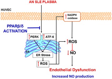
Background: We tested whether GW0742, a peroxisome proliferator-activated receptor beta/delta (PPARβ/δ) agonist, improves endothelial dysfunction induced by plasma from patients with systemic lupus erythematosus (SLE) involving the inhibition of endoplasmic reticulum (ER) stress.

Methods: A total of 12 non-pregnant women with lupus and 5 non-pregnant healthy women (controls) participated in the study. Cytokines and double-stranded DNA autoantibodies (anti-dsDNA) were tested in plasma samples. Endothelial cells, isolated from human umbilical cord veins (HUVECs), were used to measure nitric oxide (NO), intracellular reactive oxygen species (ROS) production, nicotinamide adenine dinucleotide phosphate (NADPH) oxidase activity, and ER stress markers.

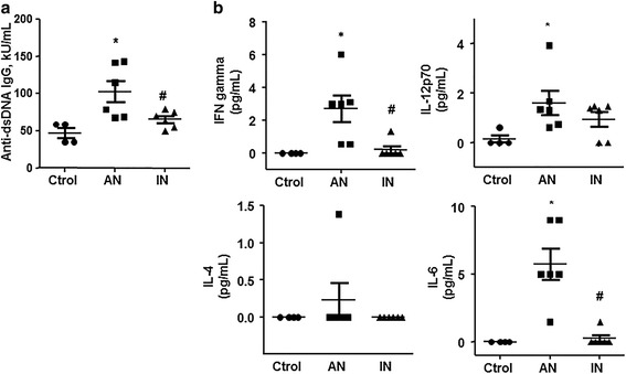
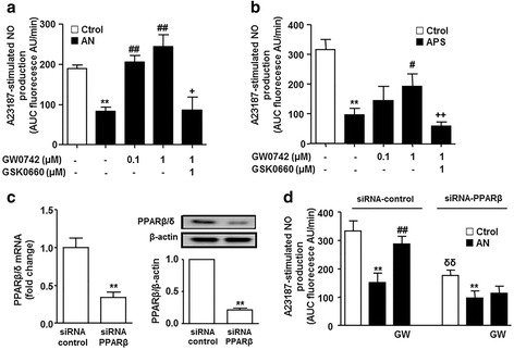
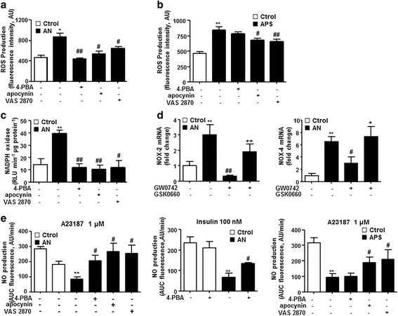
Results: Interferon-γ, interleukin-6, and interleukin-12 levels were significantly increased in plasma from patients with SLE with active nephritis (AN), as compared to both patients with SLE with inactive nephritis (IN) and the control group. The NO production stimulated by both the calcium ionophore A23187 and insulin was significantly reduced in HUVECs incubated with plasma from patients with AN-SLE as compared with the control group. Plasma from patients with IN-SLE did not modify A23187-stimulated NO production. Increased ROS production and NADPH oxidase activity were found in HUVECs incubated with plasma from patients with AN-SLE, which were suppressed by the ER stress inhibitor 4-PBA and the NADPH oxidase inhibitors, apocynin and VAS2870. GW0742 incubation restored the impaired NO production, the increased ROS levels, and the increased ER stress markers induced by plasma from patients with AN-SLE. These protective effects were abolished by the PPARβ/δ antagonist GSK0660 and by silencing PPARβ/δ.

Conclusions: PPARβ/δ activation may be an important target to control endothelial dysfunction in patients with SLE.

Keywords: Endoplasmic reticulum; Endothelial dysfunction; PPARβ/δ; Systemic lupus erythematosus.


Conflict of interest statement

Author’s information

MR is a postdoctoral fellow of the Reincorporación de Doctores del Plan Propio de Investigación, University of Granada, Spain, and MS is a postdoctoral fellow of Junta de Andalucía, Spain.

Ethics approval and consent to participate

The study was conducted in accordance with The Code of Ethics of the World Medical Association (Declaration of Helsinki). The research was also approved by the Institutional Review Board Committee at the University of Granada (Reference number: 826) and all subjects gave written informed consent.

Consent for publication

All authors provided written consent to Arthritis Research & Therapy to publish this research article.

Competing interests

The authors declare that they have no competing interests.

Publisher’s Note

Springer Nature remains neutral with regard to jurisdictional claims in published maps and institutional affiliations.

Figures

Fig. 1
Fig. 1
Autoantibodies and proinflammatory cytokine levels in plasma from participants. Anti-dsDNA (a) and interferon gamma (IFN-γ), interleukin 4 (IL-4), interleukin 6 (IL-6), and active interleukin 12 heterodimer (IL12p70) (b) were measured by a multiplex assay using luminex technology in plasma from patients with systemic lupus erythematosus (SLE) with active nephritis (AN) and with inactive nephritis (IN), and from healthy controls (Ctrol). Values are expressed as mean ± SEM (n = 5–6).* P < 0.05 vs Ctrol. # P < 0.05 vs AN-SLE
Fig. 2
Fig. 2
Effects of plasma incubation on stimulated nitric oxide (NO) production in human umbilical cord vein endothelial cells (HUVECs). NO production was stimulated by the calcium ionophore A23187 (a) or by insulin (b) in HUVECs incubated in plasma from patients with systemic lupus erythematosus (SLE) with active nephritis (AN) and with inactive nephritis (IN), and from healthy controls (Ctrol). NO release was estimated from the area under the curve (AUC) of the fluorescent signal of 4,5-diaminofluorescein (DAF-2) for 30 min in HUVECs stimulated with the drug. Values are expressed as mean ± SEM (n = 5–6). ** P < 0.01 vs Ctrol. # P < 0.05 vs AN-SLE
Fig. 3
Fig. 3
Effects of PPARβ/δ activation on endothelial nitric oxide (NO) production. NO production was stimulated by the calcium ionophore A23187 (a) in human umbilical cord vein endothelial cells (HUVECs) incubated in plasma from patients with systemic lupus erythematosus (SLE) with active nephritis (AN) or healthy controls (Ctrol), or in plasma from patients with antiphospholipid syndrome (APS) (b), in the presence and/or in absence of GW0742 and GSK0660. Values are expressed as mean ± SEM (n = 5–6). c Expression of PPARβ/δ at the level of mRNA expression by real time RT-PCR and protein by western blot in HUVECs transfected with either PPAR-β-specific siRNA (siRNA-PPAR-β) or empty vector (siRNA-control). Data are presented as gene expression normalized to Glyceraldehyde-3-phosphate dehydrogenase (GAPDH) levels or densitometric protein band and normalized to the corresponding β-actin. Results are representative of six independent experiments. ** P < 0.05 vs siRNA-control. d A23187-mediated NO production in control siRNA and siRNA-PPAR-β cells incubated in plasma from patients with AN-SLE or Ctrol for 24 h, in the presence or absence of GW0742 (1 μmol/L). All data are mean ± SEM (n = 8). NO release was estimated from the area under the curve (AUC) of the fluorescent signal of 4,5-diaminofluorescein (DAF-2) for 30 min of stimulation. ** P < 0.01 vs Ctrol. ## P < 0.01vs without PPAR agonist. + P < 0.05 vs GW0742 column. δδ P < 0.01 vs Ctrol siRNA-control column
Fig. 4
Fig. 4
Effects of PPARβ/δ activation on intracellular Reactive oxygen species (ROS) production in human umbilical cord vein endothelial cells (HUVECs). a ROS production in HUVECs incubated in plasma from patients with systemic lupus erythematosus (SLE) with active nephritis (AN) or healthy controls (Ctrol), in the presence and/or in absence of GW0742 and GSK0660. Values are expressed as mean ± SEM (n = 5–6). b ROS production in control siRNA and siRNA-PPAR-β cells incubated in plasma from patients with AN-SLE or Ctrol for 24 h, in the presence or absence of GW0742 (1 μmol/L). All data are mean ± SEM (n = 8). ROS measured by fluorescence in CM-H2DCFDA-loaded cells. * P < 0.05 and ** P < 0.01 vs Ctrol. ## P < 0.01vs without PPAR agonist. ++ P < 0.01 vs GW0742 column. δ P < 0.05 vs Ctrol siRNA-control column
Fig. 5
Fig. 5
Role of endoplasmic reticulum (ER)-stress in endothelial dysfunction induced by plasma from patients with systemic lupus erythematosus (SLE) and active nephritis (AN). Reactive oxygen species (ROS) production (a, b), measured by fluorescence in CM-H2DCFDA, and nicotinamide adenine dinucleotide phosphate (NADPH) oxidase activity (c), measured by lucigenin-enhanced chemiluminescence, measured in human umbilical cord vein endothelial cells (HUVECs) incubated in plasma from patients with SLE with AN, or in plasma from patients with antiphospholipid syndrome (APS), or healthy controls (Ctrol), in the presence and/or in absence of 4-PBA, apocynin and VAS2870. d mRNA levels of NOX2 and NOX4 in HUVECs incubated in plasma from patients with SLE with AN or Ctrol, in the presence and/or in absence of GW0742 and GSK0660. e Nitric oxide (NO) production estimated by the area under the curve (AUC) of the fluorescent signal of 4,5-diaminofluorescein (DAF-2) after stimulation with the calcium ionophore A23187 or insulin, in HUVECs incubated in plasma from patients with SLE with AN, or APS, or Ctrol, in the presence and/or in absence of 4-PBA, apocynin and VAS2870. Values are expressed as mean ± SEM (n = 5–6). * P < 0.05 and ** P < 0.01 vs Ctrol. # P < 0.05 and ## P < 0.01vs AN group without drugs. + P < 0.05 and ++ P < 0.01 vs GW0742 column
Fig. 6
Fig. 6
Effects of PPARβ/δ activation on endoplasmic reticulum (ER)-stress-induced endothelial dysfunction in human umbilical cord vein endothelial cells (HUVECs). Nitric oxide (NO) production stimulated by the calcium ionophore A23187 (a) and reactive oxygen species (ROS) production (b), measured by fluorescence in CM-H2DCFDA, in HUVECs incubated with ER-inducer tunicamycin for 24 h in the presence and/or in absence of 4-PBA, apocynin, VAS2870, GW0742, and GSK0660. Values are expressed as mean ± SEM (n = 5–6). **P < 0.01 vs healthy controls (Ctrol). #P < 0.05 and ##P < 0.01vs tunicamycin group without drugs. ++P < 0.01 vs GW0742 column
Fig. 7
Fig. 7
Effects of PPARβ/δ activation on endoplasmic reticulum (ER)-stress markers induced by incubation of plasma from patients with systemic lupus erythematosus (SLE) in human umbilical vein endothelial cells (HUVECs). Bip, IRE-1α, PERK, ATF-6, and CHOP mRNA levels, and protein expression of phospho-PERK, phospho-IRE-1α, ATF-6, and CHOP in HUVECs incubated in plasma from patients with SLE with active nephritis (AN) or healthy controls (Ctrol), in the presence and/or absence of GW0742 and GSK0660. Values are expressed as mean ± SEM (n = 5–6). * P < 0.05 and ** P < 0.01 vs Ctrol. # P < 0.05 and ## P < 0.01vs without PPAR agonist. + P < 0.05 and ++ P < 0.01 vs GW0742 column
Fig. 8
Fig. 8
Proposed mechanism of the protective effects of PPARβ/δ activation in systemic lupus erythematosus (SLE) endothelial dysfunction. SLE plasma incubation induced endothelial dysfunction in human umbilical cord vein endothelial cells (HUVECs) by sequential activation of endoplasmic reticulum (ER)-stress and nicotinamide adenine dinucleotide phosphate (NADPH) oxidase. PPARβ/δ activation inhibited both ER stress and NADPH oxidase-driven reactive oxygen species (ROS) production reducing nitric oxide (NO) inactivation. AN, active nephritis

Similar articles

Cited by

References

    1. Al-Herz A, Ensworth S, Shojania K, Esdaile JM. Cardiovascular risk factor screening in systemic lupus erythematosus. J Rheumatol. 2003;30:493–6. - PubMed
    1. Urowitz MB, Gladman D, Ibañez D, Fortin P, Sanchez-Guerrero J, Bae S, et al. Clinical manifestations and coronary artery disease risk factors at diagnosis of systemic lupus erythematosus: data from an international inception cohort. Lupus. 2007;16:731–5. doi: 10.1177/0961203307081113. - DOI - PubMed
    1. Sabio JM, Martinez-Bordonado J, Sánchez-Berná I, Vargas-Hitos JA, Mediavilla JD, Navarrete-Navarrete N, et al. Nighttime blood pressure patterns and subclinical atherosclerosis in women with systemic lupus erythematosus. J Rheumatol. 2015;42:2310–7. doi: 10.3899/jrheum.150531. - DOI - PubMed
    1. Alves JD, Ames PR. Atherosclerosis, oxidative stress and auto-antibodies in systemic lupus erythematosus and primary antiphospholipid syndrome. Immunobiology. 2003;207:23–8. doi: 10.1078/0171-2985-00215. - DOI - PubMed
    1. Bijl M. Endothelial activation, endothelial dysfunction and premature atherosclerosis in systemic autoimmune diseases. Neth J Med. 2003;61:273–7. - PubMed

MeSH terms

Substances