. 2017 Dec 19;114(51):13507-13512.
doi: 10.1073/pnas.1714386114. Epub 2017 Dec 5.

Crystal structure of human IRAK1

Affiliations

Crystal structure of human IRAK1

Li Wang et al. Proc Natl Acad Sci U S A. .

Abstract

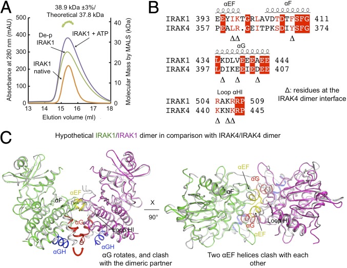
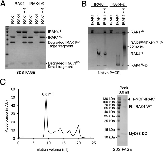
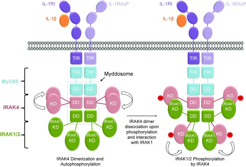
Interleukin 1 (IL-1) receptor-associated kinases (IRAKs) are serine/threonine kinases that play critical roles in initiating innate immune responses against foreign pathogens and other types of dangers through their role in Toll-like receptor (TLR) and interleukin 1 receptor (IL-1R) mediated signaling pathways. Upon ligand binding, TLRs and IL-1Rs recruit adaptor proteins, such as myeloid differentiation primary response gene 88 (MyD88), to the membrane, which in turn recruit IRAKs via the death domains in these proteins to form the Myddosome complex, leading to IRAK kinase activation. Despite their biological and clinical significance, only the IRAK4 kinase domain structure has been determined among the four IRAK family members. Here, we report the crystal structure of the human IRAK1 kinase domain in complex with a small molecule inhibitor. The structure reveals both similarities and differences between IRAK1 and IRAK4 and is suggestive of approaches to develop IRAK1- or IRAK4-specific inhibitors for potential therapeutic applications. While the IRAK4 kinase domain is capable of homodimerization in the unphosphorylated state, we found that the IRAK1 kinase domain is constitutively monomeric regardless of its phosphorylation state. Additionally, the IRAK1 kinase domain forms heterodimers with the phosphorylated, but not unphosphorylated, IRAK4 kinase domain. Collectively, these data indicate a two-step kinase activation process in which the IRAK4 kinase domain first homodimerizes in the Myddosome, leading to its trans-autophosphorylation and activation. The phosphorylated IRAK4 kinase domain then forms heterodimers with the IRAK1 kinase domain within the Myddosome, leading to its subsequent phosphorylation and activation.

Keywords: IRAK1; crystal structure; interleukin 1 receptor-associated kinases.


Conflict of interest statement

The authors declare no conflict of interest.

Figures

Fig. 1.
Fig. 1.
IRAK1 expression and purification. (A) Domain organizations of IRAK1 and IRAK4. DD, death domain; ProST, proline/serine/threonine-rich domain. (B) SDS-PAGE of the first-step Co-affinity purification. MW, molecular weight. (C) SDS-PAGE of the sample before and after His-tag removal by TEV protease treatment overnight at 4 °C. (D) Gel filtration profile of His-tag removed IRAK1 on a Superdex 75 10/300 column and the corresponding SDS-PAGE of the elution fractions. The degraded IRAK1 fragments coeluted with the nondegraded construct band.
Fig. 2.
Fig. 2.
Proteolytic screening of the IRAK1 kinase domain construct. (A) SDS-PAGE of purified IRAK1 treated with various proteases at a 1,000:1 (wt/wt) ratio at 37 °C for 1 h. (B) SDS-PAGE of purified IRAK1 before clostripain treatment and IRAK1 from washed crystals.
Fig. 3.
Fig. 3.
Structure of inhibitor-bound IRAK1. (A) Chemical structure of the dual IRAK1/4 inhibitor JH-I-25 used in the cocrystallization. (B) A ribbon diagram of IRAK1 in complex with the inhibitor. The secondary structures are labeled, and the JH-I-25 inhibitor is shown with carbon atoms in magenta, nitrogen atoms in blue, and oxygen atoms in red, superimposed with its electron density in gray. IRAK1 is in green with the exception of the long, gray-colored IRAK1-specific insertion. (C) Superposition between IRAK1 (green) and IRAK4 (pink, PDB ID code 2NRU). The main differences between these two kinase domains at the αBβ1 loop, the αDE loop, and the αGH insertion are labeled. The different locations of C302 of IRAK1 and its sequence-conserved counterpart C276 of IRAK4 are indicated. (D) The regulatory spine and the K239-E259 salt bridge both indicate an active conformation of IRAK1. (E) Sequence alignment between IRAK1 and IRAK4 at four regions with significantly different structures.
Fig. 4.
Fig. 4.
Detailed mode of inhibitor interaction. (A) A hydrogen-bonding network around the IRAK1 gatekeeper residue Y288 near the head of the inhibitor. (B) Interactions of IRAK1 with JH-I-25. IRAK1 residues are labeled in green while the corresponding IRAK4 residues are shown in pink. (C) Surface diagrams of IRAK1 (Left, green) and IRAK4 (Right, pink) at the tail of the inhibitor pocket, showing the openness of IRAK1 and closeness of IRAK4. (D) Superimposed IRAK1 and IRAK4 at the region of the ATP front pocket showing that an insertion in IRAK4 and distinct loop conformations are responsible for the difference in the openness of this region. (E) Side chain directions of C302 and C307 in IRAK1 may allow design of covalent inhibitors, but the side chain direction of C276 of IRAK4 may not.
Fig. 5.
Fig. 5.
IRAK1 kinase domain is a monomer in solution in both phosphorylated and unphosphorylated states. (A) Gel filtration profiles and multiangle light scattering measurement showed that the IRAK1 kinase domain is a monomer in different states, different from the dimeric state of the IRAK4 kinase domain in the unphosphorylated form. The green track on the top marks the measured molecular mass of the dephosphorylated IRAK1 kinase domain. mAU, absorption units. (B) Sequence alignment between IRAK1 and IRAK4 at the IRAK4 dimerization interface. Important IRAK4 dimerization residues are indicated by triangles. (C) A model of a hypothetical IRAK1 homodimer would have caused clash in several dimerization elements including the αG and the αEF regions.
Fig. 6.
Fig. 6.
IRAK1 interacts weakly with phosphorylated IRAK4. (A) IRAK1 and two forms of IRAK4, unphosphorylated (left lanes, “IRAK4”) and phosphorylated (shown as an encircled P; right lanes, “IRAK4-P”) on an SDS-PAGE. (B) IRAK1 and two forms of IRAK4, unphosphorylated (left lanes, “IRAK4”) and phosphorylated (right lanes, “IRAK4-P”) on a native PAGE, showing the shifted band containing IRAK1 and phosphorylated IRAK4. (C) Gel filtration profile (Left) and SDS-PAGE (Right) of the peak fraction for the reconstituted complex of the MyD88 death domain (DD), full-length (FL) IRAK4, and His-MBP–tagged IRAK1 containing the DD and the kinase domain.
Fig. 7.
Fig. 7.
A schematic diagram of IRAK activation within the Myddosome shown here for the IL-1 receptors. Upon ligand binding, IL-1 receptor (IL-1R) and IL-1 receptor accessary protein (IL-1RAcp) recruit MyD88, which in turn recruits the upstream kinase IRAK4 and then the downstream kinase IRAK1 or IRAK2. IRAK4 recruitment leads to increased local concentration of the kinase domain, leading to its dimerization and trans-autophosphorylation. Phosphorylated IRAK4 dissociates from the dimer to interact with and phosphorylate IRAK1 or IRAK2, leading to the first step of its activation.

Similar articles

Cited by

References

    1. Cao Z, Henzel WJ, Gao X. IRAK: A kinase associated with the interleukin-1 receptor. Science. 1996;271:1128–1131. - PubMed
    1. Muzio M, Ni J, Feng P, Dixit VM. IRAK (Pelle) family member IRAK-2 and MyD88 as proximal mediators of IL-1 signaling. Science. 1997;278:1612–1615. - PubMed
    1. Wesche H, et al. IRAK-M is a novel member of the Pelle/interleukin-1 receptor-associated kinase (IRAK) family. J Biol Chem. 1999;274:19403–19410. - PubMed
    1. Li S, Strelow A, Fontana EJ, Wesche H. IRAK-4: A novel member of the IRAK family with the properties of an IRAK-kinase. Proc Natl Acad Sci USA. 2002;99:5567–5572. - PMC - PubMed
    1. Ye H, et al. Distinct molecular mechanism for initiating TRAF6 signalling. Nature. 2002;418:443–447. - PubMed

Publication types

MeSH terms

Substances

Associated data

LinkOut - more resources