. 2017 Dec 5;8(1):1956.
doi: 10.1038/s41467-017-02012-2.

Mechanistic insight into TRIP13-catalyzed Mad2 structural transition and spindle checkpoint silencing

Affiliations

Mechanistic insight into TRIP13-catalyzed Mad2 structural transition and spindle checkpoint silencing

Melissa L Brulotte et al. Nat Commun. .

Abstract

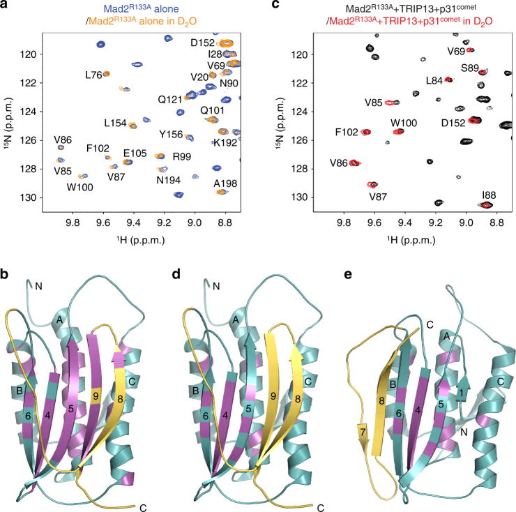
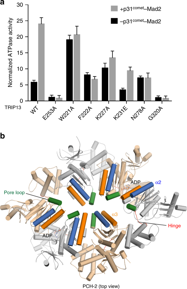
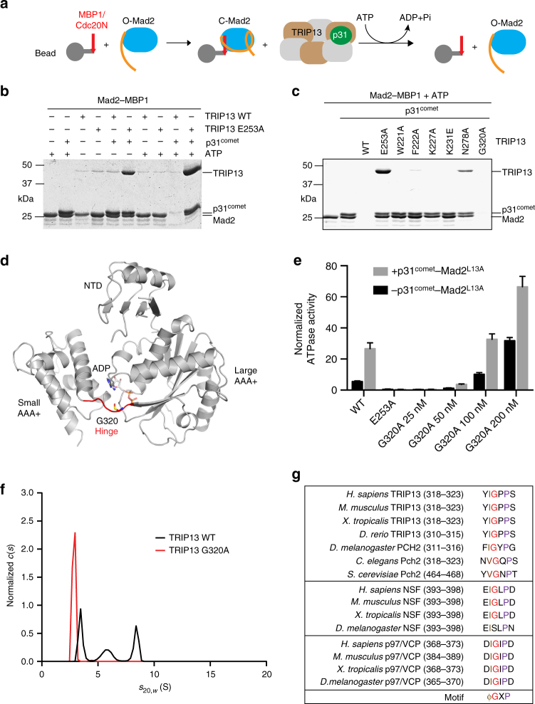
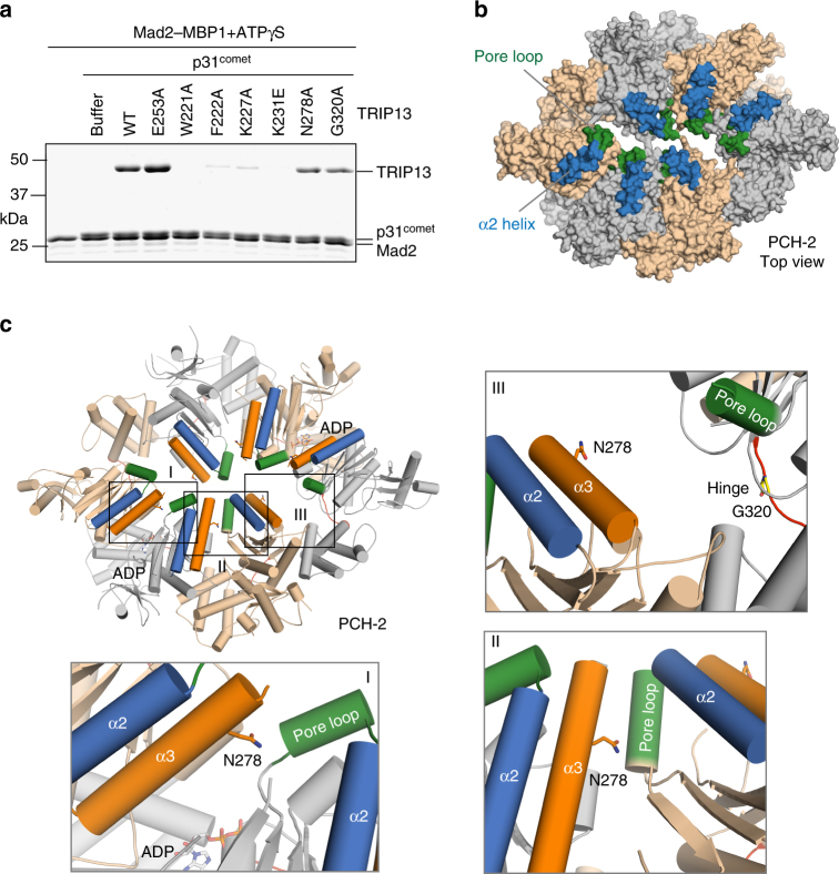
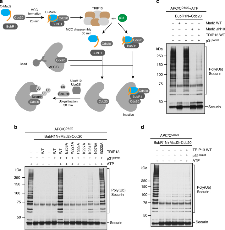
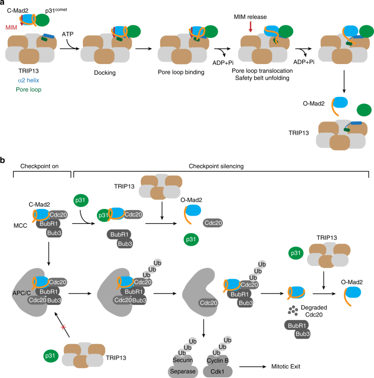
The spindle checkpoint maintains genomic stability and prevents aneuploidy. Unattached kinetochores convert the latent open conformer of the checkpoint protein Mad2 (O-Mad2) to the active closed conformer (C-Mad2), bound to Cdc20. C-Mad2-Cdc20 is incorporated into the mitotic checkpoint complex (MCC), which inhibits the anaphase-promoting complex/cyclosome (APC/C). The C-Mad2-binding protein p31comet and the ATPase TRIP13 promote MCC disassembly and checkpoint silencing. Here, using nuclear magnetic resonance (NMR) spectroscopy, we show that TRIP13 and p31comet catalyze the conversion of C-Mad2 to O-Mad2, without disrupting its stably folded core. We determine the crystal structure of human TRIP13, and identify functional TRIP13 residues that mediate p31comet-Mad2 binding and couple ATP hydrolysis to local unfolding of Mad2. TRIP13 and p31comet prevent APC/C inhibition by MCC components, but cannot reactivate APC/C already bound to MCC. Therefore, TRIP13-p31comet intercepts and disassembles free MCC not bound to APC/C through mediating the local unfolding of the Mad2 C-terminal region.


Conflict of interest statement

The authors declare no competing financial interests.

Figures

Fig. 1
Fig. 1
TRIP13 catalyzes C-Mad2 to O-Mad2 conversion in the presence of p31comet. a Ribbon diagrams of O-Mad2 (left), unliganded C-Mad2 (middle), and C-Mad2 bound to its high-affinity artificial ligand MBP1 (right). The C-terminal region that undergoes a large conformational change and forms the safety belt structure in C-Mad2 is colored yellow while the rest of the protein is in cyan. Strands in the major β sheet are labeled with their numbers. The N- and C-termini are indicated. All protein structure figures in this study were generated with PyMOL (Schrödinger, LLC; http://www.pymol.org). b Overlay of regions of the 1H–15N HSQC spectra of 15N-C-Mad2R133A (in magenta) and 15N-Mad2R133A before the addition of TRIP13 and ΔN35-p31comet (in blue). c Overlay of regions of the 1H–15N HSQC spectra of 15N-O-Mad2R133A (in cyan) and 15N-Mad2R133A after the addition of ATP and sub-stoichiometric amounts of TRIP13 and ΔN35-p31comet (in black)
Fig. 2
Fig. 2
TRIP13-catalyzed structural transition of Mad2 does not involve Mad2 global unfolding. a Overlay of regions of the 1H–15N HSQC spectra of 15N-labeled C-Mad2R133A in H2O (blue) and C-Mad2R133A in D2O (orange). Residues with slow-exchanging amides are labeled. b Ribbon diagram of unliganded C-Mad2, with slow-exchanging residues in the absence of TRIP13 colored magenta. Strands in the major β sheet are labeled with their numbers. The N- and C-termini are indicated. c Overlay of regions of the 1H–15N HSQC spectra of 15N-labeled Mad2R133A in H2O (black) and D2O (red) after the addition of ATP and sub-stoichiometric amounts of TRIP13 and ΔN35-p31comet. Residues with slow-exchanging amides are labeled. d Ribbon diagram of unliganded C-Mad2, with slow-exchanging residues in the presence of TRIP13 colored magenta. e Ribbon diagram of O-Mad2, with slow-exchanging residues in the presence of TRIP13 colored magenta
Fig. 3
Fig. 3
Crystal structure of human TRIP13 in its ADP-bound state. a Surface and ribbon diagrams of C. elegans PCH-2 (PDB ID: 4XGU) in top and side views. Neighboring protomers are shown in different colors. b Ribbon diagram of the crystal structure of human TRIP13, with the bound ADP shown in sticks. c Overlay of the ribbon diagrams of the structures of human TRIP13 (orange) and the ADP-bound protomer of C. elegans PCH-2 (gray; except the pore loop, which is colored green), with the bound ADP molecules shown in sticks
Fig. 4
Fig. 4
Identification of TRIP13 mutants with defective p31comet–Mad2 stimulation. a Normalized ATPase activities of the indicated TRIP13 proteins at 25 nM with (+) or without (−) 50 nM ΔN35-p31comet–Mad2L13A. Mean ± SD; n = 9. b Cartoon diagram of C. elegans PCH-2 in the top view. The pore loop, the α2 helix, the α3 helix, and the hinge are shown in green, blue, orange, and red, respectively. The bound ADP molecules are shown in sticks
Fig. 5
Fig. 5
The TRIP13 hinge is required for conformational dynamics and oligomerization. a Schematic drawing of the Mad2 dissociation assay. b MBP1-coupled beads were first incubated with Mad2 and then incubated with or without TRIP13 wild type (WT) or E253A (1 µM), ΔN35-p31comet (2 µM), or ATP (1 mM). Proteins bound to beads were analyzed by SDS-PAGE and stained with Coomassie blue. c MBP1-coupled beads were first incubated with Mad2 and then incubated with TRIP13 wild type (WT) and the indicated mutants (500 nM) in the presence of ΔN35-p31comet (1 µM) and ATP (1 mM). Proteins bound to beads were analyzed by SDS-PAGE and stained with Coomassie blue. d Ribbon diagram of human TRIP13, with the hinge connecting the large and small AAA+ subdomains colored red. ADP and G320 are shown in sticks. e ATPase activities of TRIP13 WT at 25 nM and G320A at the indicated concentrations with (+) or without (−) ΔN35-p31comet–Mad2L13A. The concentrations of p31comet–Mad2 were kept at 2:1 molar ratio of those of TRIP13. Mean ± SD; n = 8. f Sedimentation velocity analysis of TRIP13 WT and G320A in the absence of ATP. g Sequence alignment of the hinge region of TRIP13, the D1 domain NSF, and the D1 domain p97/VCP from different organisms
Fig. 6
Fig. 6
Identification of a p31comet–Mad2-binding site on TRIP13. a MBP1-coupled beads were first incubated with Mad2, and then incubated with TRIP13 wild type (WT) or the indicated mutants (500 nM), ΔN35-p31comet (1 µM), and ATPγS (1 mM). Proteins bound to beads were analyzed by SDS-PAGE and stained with Coomassie blue. b Surface diagram of C. elegans PCH-2 in the top view, with the pore loop and α2 helix colored green and blue, respectively. c Cartoon diagram of C. elegans PCH-2, with zoomed-in views of the boxed interfaces (I–III) shown. N278, ADP, and G320 are shown in sticks. The pore loop, the α2 helix, and the α3 helix are colored green, blue, and orange, respectively. Each of the three types of interfaces is present twice in the PCH-2 hexamer
Fig. 7
Fig. 7
TRIP13 prevents APC/CCdc20 inhibition by MCC components. a Schematic diagram of the APC/C ubiquitination assay in the absence or presence of MCC components, TRIP13, or p31comet. b Ubiquitination of securin-Myc by APC/CCdc20 in the presence of MCC components (780 nM BubR1N, 1 μM Mad2, and 600 nM Cdc20), TRIP13 wild type (WT) or the indicated mutants (50 nM), and ΔN35-p31comet (1 µM). MCC components were pre-incubated with TRIP13, p31comet, and ATP (1 mM). The protein mixture was then added to APC/CCdc20. The ubiquitination reaction mixtures were blotted with the anti-Myc antibody. c Same as in b except that Mad2 ΔN10 and only TRIP13 WT were used. d Ubiquitination of securin-Myc by APC/CCdc20 that was first incubated with MCC components (780 nM BubR1N, 1 μM Mad2, and 600 nM Cdc20) for 20 min and then incubated with TRIP13 WT (50 nM) and ΔN35-p31comet (1 µM). The ubiquitination reaction mixtures were blotted with the anti-Myc antibody
Fig. 8
Fig. 8
Mechanisms of TRIP13-dependent Mad2 conformational change and checkpoint silencing. a Model of TRIP13-catalyzed conversion of C-Mad2 to O-Mad2. The p31comet–C-Mad2–MIM complex docks on the α2 helix of TRIP13. The pore loop of TRIP13 engages the C-terminus of Mad2. ATP hydrolysis drives the translocation of the pore loop and the associated Mad2 C-terminus, leading to the local unfolding of the C-terminal safety belt of C-Mad2. This Mad2 local unfolding releases MIM and disrupts the p31comet–Mad2 interaction. Mad2 refolding produces O-Mad2. Both p31comet and O-Mad2 dissociate from TRIP13. b Model of TRIP13-dependent checkpoint silencing. Upon spindle checkpoint activation, MCC is produced at unattached kinetochores and diffuses into the cytosol to inhibit APC/CCdc20. During checkpoint silencing, the production of MCC is attenuated. p31comet binds to C-Mad2 in MCC and displaces BubR1–Bub3 from MCC. TRIP13 then binds to p31comet–C-Mad2–Cdc20, disassembles the C-Mad2–Cdc20 complex, and converts C-Mad2 to O-Mad2. Because TRIP13 cannot act on MCC already bound to APC/CCdc20, MCC must first be released from APC/CCdc20 through alternative mechanisms. Cdc20 ubiquitination is a potential MCC-releasing mechanism. Proteasome-mediated degradation of ubiquitinated Cdc20 then disassembles MCC. C-Mad2 that persists during this process can be recognized by p31comet and TRIP13, and converted to O-Mad2. Collectively, these mechanisms reduce the levels of MCC and promote the activation of APC/CCdc20, which ubiquitinates securin and cyclin B1 to trigger chromosome segregation and mitotic exit

References

    1. Jia L, Kim S, Yu H. Tracking spindle checkpoint signals from kinetochores to APC/C. Trends Biochem. Sci. 2013;38:302–311. doi: 10.1016/j.tibs.2013.03.004. - DOI - PubMed
    1. London N, Biggins S. Signalling dynamics in the spindle checkpoint response. Nat. Rev. Mol. Cell Biol. 2014;15:736–747. doi: 10.1038/nrm3888. - DOI - PMC - PubMed
    1. Musacchio A. The molecular biology of spindle assembly checkpoint signaling dynamics. Curr. Biol. 2015;25:R1002–R1018. doi: 10.1016/j.cub.2015.08.051. - DOI - PubMed
    1. Yu H. Cdc20: a WD40 activator for a cell cycle degradation machine. Mol. Cell. 2007;27:3–16. doi: 10.1016/j.molcel.2007.06.009. - DOI - PubMed
    1. Kim S, Yu H. Mutual regulation between the spindle checkpoint and APC/C. Semin. Cell Dev. Biol. 2011;22:551–558. doi: 10.1016/j.semcdb.2011.03.008. - DOI - PMC - PubMed

Publication types

MeSH terms

Substances