. 2017 Dec 5;8(1):1941.
doi: 10.1038/s41467-017-02164-1.

Polη O-GlcNAcylation governs genome integrity during translesion DNA synthesis

Affiliations

Polη O-GlcNAcylation governs genome integrity during translesion DNA synthesis

Xiaolu Ma et al. Nat Commun. .

Abstract

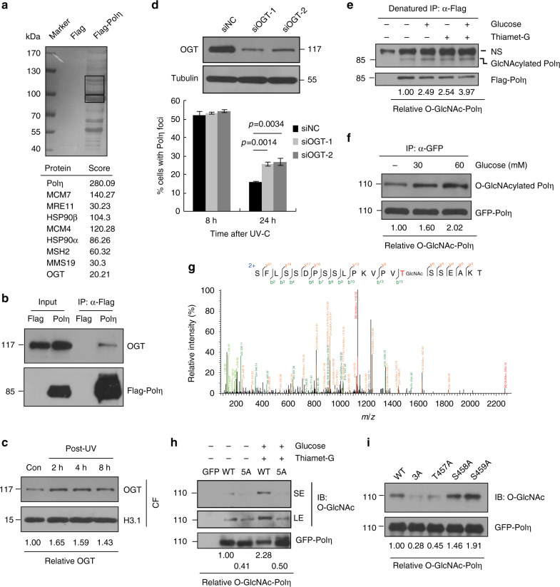
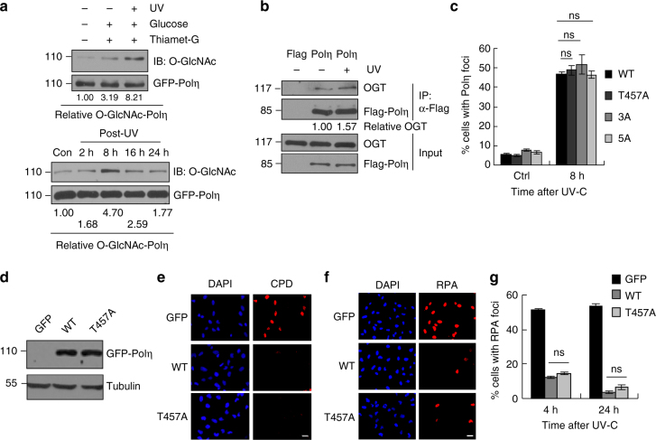
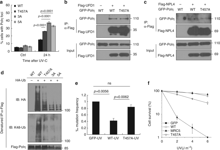
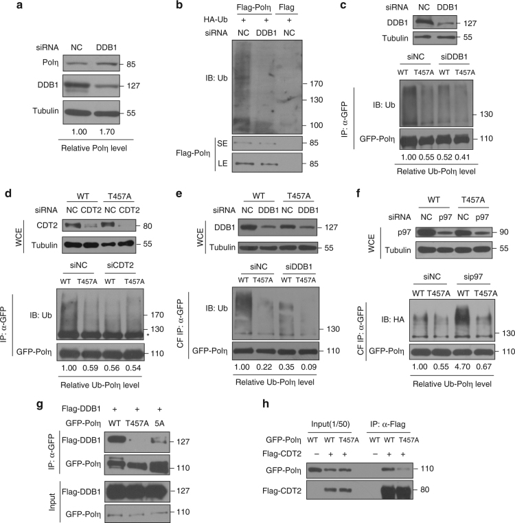
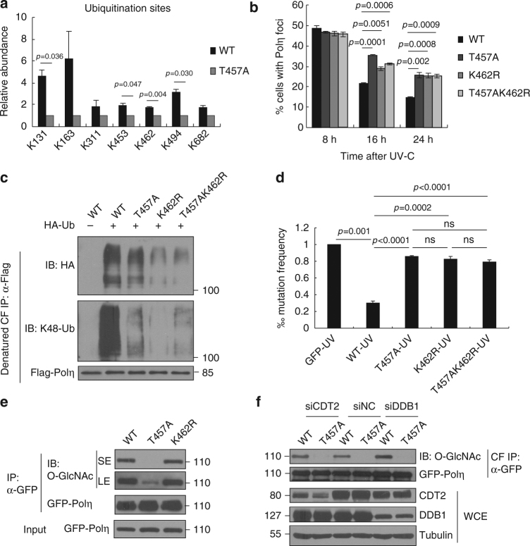
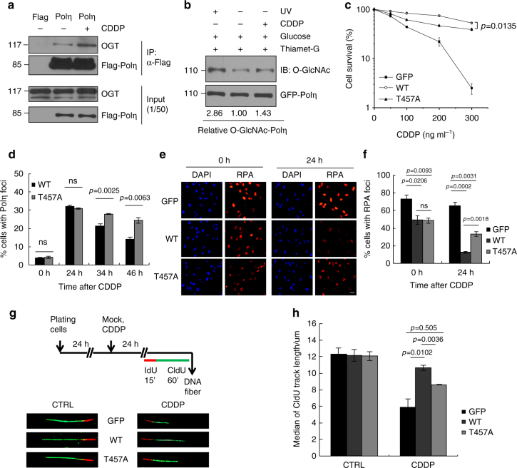
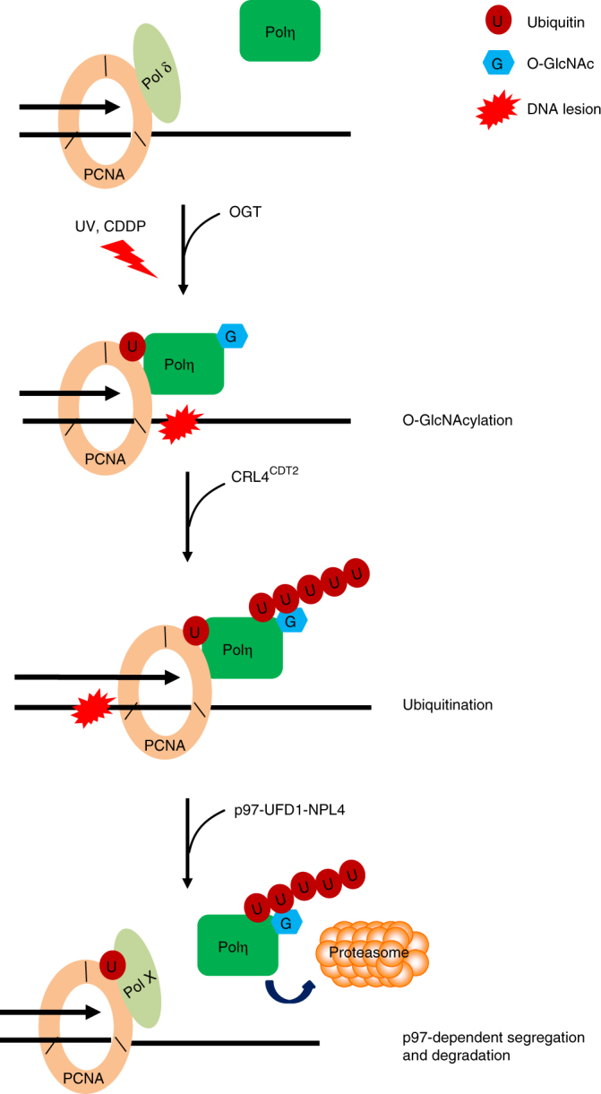
DNA polymerase η (Polη) facilitates translesion DNA synthesis (TLS) across ultraviolet (UV) irradiation- and cisplatin-induced DNA lesions implicated in skin carcinogenesis and chemoresistant phenotype formation, respectively. However, whether post-translational modifications of Polη are involved in these processes remains largely unknown. Here, we reported that human Polη undergoes O-GlcNAcylation at threonine 457 by O-GlcNAc transferase upon DNA damage. Abrogation of this modification results in a reduced level of CRL4CDT2-dependent Polη polyubiquitination at lysine 462, a delayed p97-dependent removal of Polη from replication forks, and significantly enhanced UV-induced mutagenesis even though Polη focus formation and its efficacy to bypass across cyclobutane pyrimidine dimers after UV irradiation are not affected. Furthermore, the O-GlcNAc-deficient T457A mutation impairs TLS to bypass across cisplatin-induced lesions, causing increased cellular sensitivity to cisplatin. Our findings demonstrate a novel role of Polη O-GlcNAcylation in TLS regulation and genome stability maintenance and establish a new rationale to improve chemotherapeutic treatment.


Conflict of interest statement

The authors declare no competing financial interests.

Figures

Fig. 1
Fig. 1
Polη interacts with OGT and is subject to O-GlcNAcylation predominantly at T457. a The nuclear extracts of 293T cells expressing Flag-Polη were immunoprecipitated. The indicated bands (black rectangles) were cut and analyzed via mass spectrometric analysis. b 293T cell lysates expressing Flag-Polη were immunoprecipitated and detected with anti-OGT and anti-Flag antibodies. The input included 2% of the cell lysate used. c U2OS cells were irradiated with UVC (15 J m−2) and harvested at different time points later. The chromatin fractions were extracted followed by immunoblotting with OGT and H3.1 antibodies. d U2OS cells were transfected with siOGT or siNC oligos. The OGT protein level was detected by western blot. The knockdown cells were transfected with GFP-Polη, irradiated with UVC (15 J m−2) and further incubated for 24 h. The proportions of GFP-Polη-expressing cells with >30 foci were determined. Data represent means ± SEM from three independent experiments. e 293T cells transfected with Flag-Polη or empty vector were incubated with Thiamet-G and glucose. The cell lysates were denatured and immunoprecipitated with anti-Flag M2 beads followed by immunoblotting with O-GlcNAc and Flag antibodies. NS: non-specific band. f 293T cells transfected with GFP-Polη were treated with Thiamet-G and different concentrations of glucose. The cell lysates were immunoprecipitated with GFP-Trap A followed by immunoblotting with O-GlcNAc and GFP antibodies. g 293T cells transfected with Flag-Polη or empty vector were treated with Thiamet-G and glucose. The cell lysates were immunoprecipitated with anti-Flag agarose followed by HCD-MS analysis. The tryptic peptide containing a HexNAc (+203.08 Da) was detected. The b- and y-type product ions were marked on the spectrum and also illustrated along the peptide sequence shown on top of the spectrum. h, i 293T cells transfected with the indicated GFP-Polη constructs were treated as in g. The cell lysates were immunoprecipitated with GFP-Trap A followed by immunoblotting with O-GlcNAc and GFP antibodies. SE short exposure, LE long exposure
Fig. 2
Fig. 2
T457A has no effect on the efficiency of Polη to bypass CPDs. a 293T cells transfected with GFP-Polη were treated with Thiamet-G and glucose. Cells were irradiated with UVC (15 J m−2) and harvested 4 h later (top panel) or at the indicated time points (bottom panel). The cell lysates were immunoprecipitated with GFP-Trap A followed by immunoblotting with O-GlcNAc and GFP antibodies. Experiment was repeated three times. SE short exposure, LE long exposure. b 293T cells transfected with Flag-Polη were irradiated with UVC (15 J m−2) and harvested 4 h later. The cell lysates were immunoprecipitated and the relative IPed OGT was shown. Experiment was repeated twice. c U2OS cells transfected with indicated GFP-Polη constructs were UVC irradiated and incubated for 8 h. The proportions of GFP-Polη expressing cells with >30 foci were determined by counting at least 200 cells in each experiment. d Polη-deficient XP30RO cells infected with GFP, WT or T457A GFP-Polη lentivirus were selected by flow cytometry. The levels of Polη protein in stable clones were examined with anti-Polη. Tubulin: loading control. e Representative images of cells stained with CPDs (red) in ssDNA of nuclei (DAPI, blue) after UV irradiation. XP30RO cells stably expressing GFP, WT or T457A GFP-Polη were irradiated with 5 J m−2 UVC and cultured for 4 h. The cells were treated with 1% Triton-X100 for 2 min prior to fixation. Scale bars: 10 μm. f Representative images of cells stained with DAPI and RPA2 after UV irradiation. XP30RO cells stably expressing GFP, WT or T457A GFP-Polη were irradiated with 5 J m−2 UVC and further cultured. At the indicated time points, cells were treated with 0.5% Triton-X100 for 30 min followed by immunofluorescence. Scale bars: 10 μm. g Quantification of the percentage of cells with >10 RPA foci. For each cell line at each time point, at least 250 cells were counted. Data represent means ± SEM from three independent experiments. ns not significant
Fig. 3
Fig. 3
T457A impairs Polη removal from chromatin after UV irradiation. a U2OS cells transfected with WT or mutated GFP-Polη constructs were irradiated with UVC (15 J m−2) and further incubated for 24 h. The proportions of GFP-Polη-expressing cells with >30 foci were determined. Data represent means ± SEM from three independent experiments. b, c Flag-UFD1 b or Flag-NPL4 c and GFP-Polη (WT or T457A) were transfected into 293T cells. The lysates were immunoprecipitated using anti-Flag M2 beads. The inputs and immunoprecipitates were examined via western blotting using anti-GFP and anti-Flag antibodies. d 293T cells were transfected with WT or mutant Flag-Polη as well as HA-Ub or HA empty vector. The cell lysates were immunoprecipitated with anti-Flag M2 beads under denaturing condition, followed by immunoblotting with anti-HA, anti-K48-Ubiquitin, and anti-Flag antibodies. e Mutation frequency in damaged (400 J m−2 UVC) supF plasmid was determined. Data represent means ± SEM from three independent experiments. ns not significant. f XP30RO cells stably expressing GFP, WT or T457A GFP-Polη and MRC5 cells were irradiated with the indicated doses of UVC and further incubated in medium supplemented with 0.4 mM caffeine for 7–10 days. The number of cell clones was determined. Surviving fraction was expressed as a percentage of mock-treated cells. Experiment was repeated three times, giving similar results. The representative curve is shown. Error bar: s.d., n = 3
Fig. 4
Fig. 4
T457A impairs CRL4CDT2-mediated Polη polyubiqitination. a 293T cells were transfected with siDDB1 or siNC oligos. Seventy-two hours later, the cells were harvested and the protein levels of Polη and DDB1 were detected by western blotting. Tubulin: loading control. b Flag empty vector or Flag-Polη and HA-Ub were transfected in siNC- or siDDB1-treated 293T cells. The cells lysates were immunoprecipitated by anti-Flag M2 beads and analyzed by immunoblotting with anti-Ubiquitin and anti-Flag antibodies. SE short exposure, LE long exposure. c, d WT or T457A GFP-Polη and HA-Ub were transfected into siDDB1-treated c or siCDT2-treated d 293T cells. The lysates were immunoprecipitated using GFP-Trap A. The immunoprecipitates were analyzed via western blotting using anti-GFP and anti-Ubiquitin antibodies. The protein levels of DDB1 and CDT2 were detected by immunoblotting. WCE whole cell lysate. Tubulin: loading control. Asterisk denote non-specific signal. e WT or T457A GFP-Polη and HA-Ub were transfected into siDDB1-treated 293T cells. The chromatin fractions (CF) were harvested followed by immunoprecipitation with GFP-Trap A. The immunoprecipitates were analyzed as in c. f WT or T457A GFP-Polη and HA-Ub were transfected into p97 siRNA (sip97)-treated 293T cells. The chromatin fractions were immunoprecipitated and analyzed as in c. The p97 protein level was detected by western blot. g Flag-DDB1 and GFP-Polη (WT, T457A, or 5A) were transfected into 293T cells. The lysates were immunoprecipitated using GFP-Trap A. The immunoprecipitates and inputs were examined via western blotting using anti-GFP and anti-Flag antibodies. h Flag empty vector or Flag-CDT2 and GFP-Polη (WT or T457A) were transfected into 293T cells. The lysates were immunoprecipitated and analyzed as in g
Fig. 5
Fig. 5
T457A impairs Polη polyubiqitination at K462. a 293T cells transfected with Flag empty vector, WT, or T457A Flag-Polη and HA-Ub were immunoprecipitated with anti-Flag agarose in a denatured condition followed by quantitative MS analysis as described in “Methods” section. All ubiquitination sites identified in WT and T457A Polη. Error bars: s.e.m. b U2OS cells transfected with WT or mutated (T457A, K462R, and T457AK462R) GFP-Polη were irradiated by UVC (15 J m−2) and further incubated for 24 h. The proportion of GFP-Polη-expressing cells with >30 foci was determined. Data represent means ± SEM from three independent experiments. c 293 T cells were transfected with WT or mutated (T457A, K462R, and T457A/K462R) Flag-Polη and HA-Ub. The collected chromatin fractions (CFs) were denatured followed by immunoprecipitation with anti-Flag agarose. The immunoprecipitates were immunoblotted with anti-HA, anti-K48-Ubiquitin, and anti-Flag antibodies. d Mutation frequency in damaged (400 J m−2 UVC) supF plasmid was determined. Data represent means ± SEM from three independent experiments. ns not significant. e WT, T457A, or K462R GFP-Polη were transfected into 293T cells. The cell lysates were immunoprecipitated with GFP-Trap A, followed by immunoblotting with anti-O-GlcNAc and anti-GFP antibodies. “SE” and “LE” represent short and long exposure, respectively. f WT or T457A GFP-Polη were transfected into siDDB1-, siCDT2-, or siNC-treated 293T cells. The chromatin fractions were immunoprecipitated with GFP-Trap A and analyzed via western blotting with anti-O-GlcNAc and anti-GFP antibodies
Fig. 6
Fig. 6
T457 affects Polη TLS efficiency after cisplatin treatment. a 293T cells transfected with Flag-Polη were treated with CDDP (10 µg ml−1) for 2 h and changed to drug-free media for 2 h. The cell lysates were harvested for immunoprecipitation assay as in Fig. 1e. b 293T cells transfected with GFP-Polη were treated with Thiamet-G and glucose. Cells were treated with UVC (15 J m−2) irradiation or cisplatin (10 µg ml−1) and harvested at 4 or 2 h later, respectively. The cell lysates were immunoprecipitated with GFP-Trap A and analyzed with anti-O-GlcNAc and anti-GFP antibodies. c XP30RO cells stably expressing GFP, WT, or T457A GFP-Polη were treated with cisplatin for 24 h and further incubated for 7–10 days. Cell survival was analyzed as in Fig. 3f. d U2OS cells transfected with WT or T457A GFP-Polη were treated with cisplatin (5 µg ml−1) for 2 h followed by incubation with fresh media. At the indicated time points, the proportion of GFP-Polη expressing cells with >30 foci was determined. e Representative images of cells stained with DAPI and RPA2 after cisplatin treatment. XP30RO cells stably expressing GFP, WT, or T457A GFP-Polη were treated with cisplatin for 24 h and further cultured. At the indicated time points, cells were immunostained with anti-RPA2 antibody. Scale bars: 10 μm. f Quantification of the percentage of cells with >10 RPA2 foci. g Scheme of experimental design for replication fork progression in XP30RO cells stably expressing GFP, WT, or T457A GFP-Polη. After cisplatin (1 µg ml−1) treatment for 24 h, representative DNA fibers were shown. h Median lengths of nascent replication tracts (labled with CIdU) were given. Data represent means ± SEM from three independent experiments. ns not significant
Fig. 7
Fig. 7
A proposed model depicting the role of Polη O-GlcNAcylation in its removal after TLS. Upon UV or CDDP exposure, Polη is recruited to stalled replication forks and gets O-GlcNAcylated by OGT. Once Polη’s role in TLS is completed, O-GlcNAcylated Polη is ubiquitinated by CRL4CDT2 E3 ligase complex. The polyubiquitinated Polη is then recognized by p97-UFD-NPL4 complex, resulting in its dissociation from replication forks and degradation

References

    1. Johnson RE, Prakash S, Prakash L. Efficient bypass of a thymine-thymine dimer by yeast DNA polymerase, Poleta. Science. 1999;283:1001–1004. doi: 10.1126/science.283.5404.1001. - DOI - PubMed
    1. Friedberg, E. C. et al. DNA Repair and Mutagenesis, 2nd edn. (American Society for Microbiology, Washington, DC, 2005).
    1. Masutani C, et al. The XPV (xeroderma pigmentosum variant) gene encodes human DNA polymerase eta. Nature. 1999;399:700–704. doi: 10.1038/21447. - DOI - PubMed
    1. Yang W. An overview of Y-Family DNA polymerases and a case study of human DNA polymerase eta. Biochemistry. 2014;53:2793–2803. doi: 10.1021/bi500019s. - DOI - PMC - PubMed
    1. Kannouche P, et al. Domain structure, localization, and function of DNA polymerase eta, defective in xeroderma pigmentosum variant cells. Genes Dev. 2001;15:158–172. doi: 10.1101/gad.187501. - DOI - PMC - PubMed

Publication types