. 2017 Nov 2:11:341.
doi: 10.3389/fncel.2017.00341. eCollection 2017.

Noradrenaline Modulates the Membrane Potential and Holding Current of Medial Prefrontal Cortex Pyramidal Neurons via β1-Adrenergic Receptors and HCN Channels

Affiliations

Noradrenaline Modulates the Membrane Potential and Holding Current of Medial Prefrontal Cortex Pyramidal Neurons via β1-Adrenergic Receptors and HCN Channels

Katarzyna Grzelka et al. Front Cell Neurosci. .

Abstract

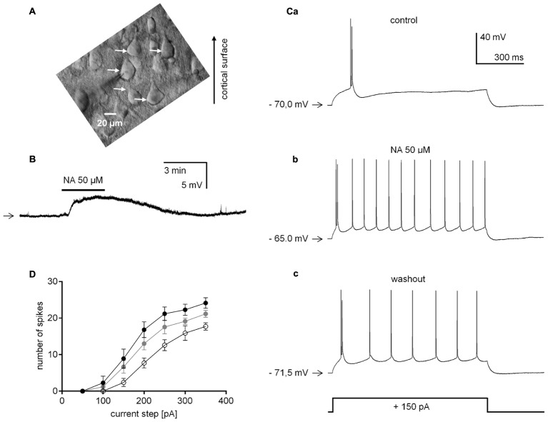
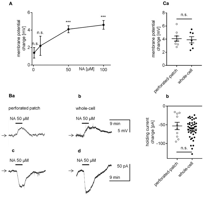
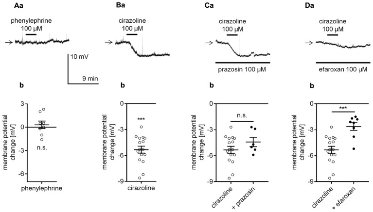
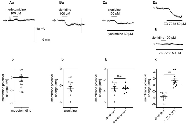
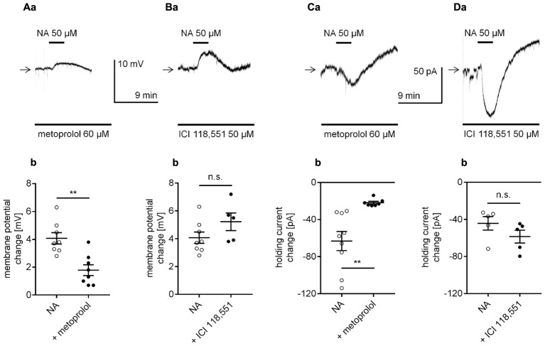
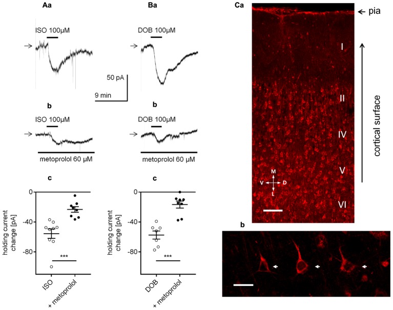
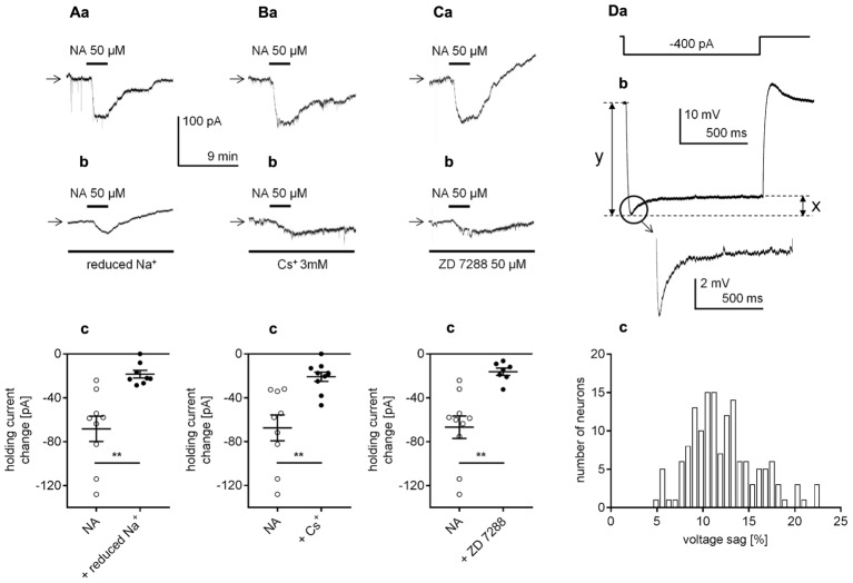
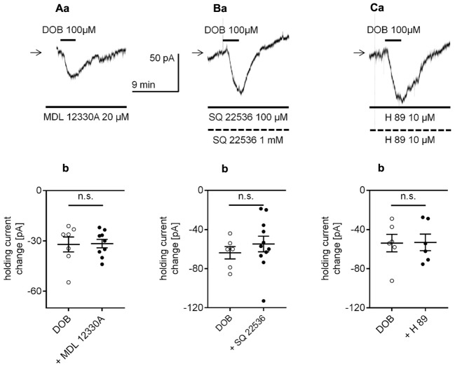
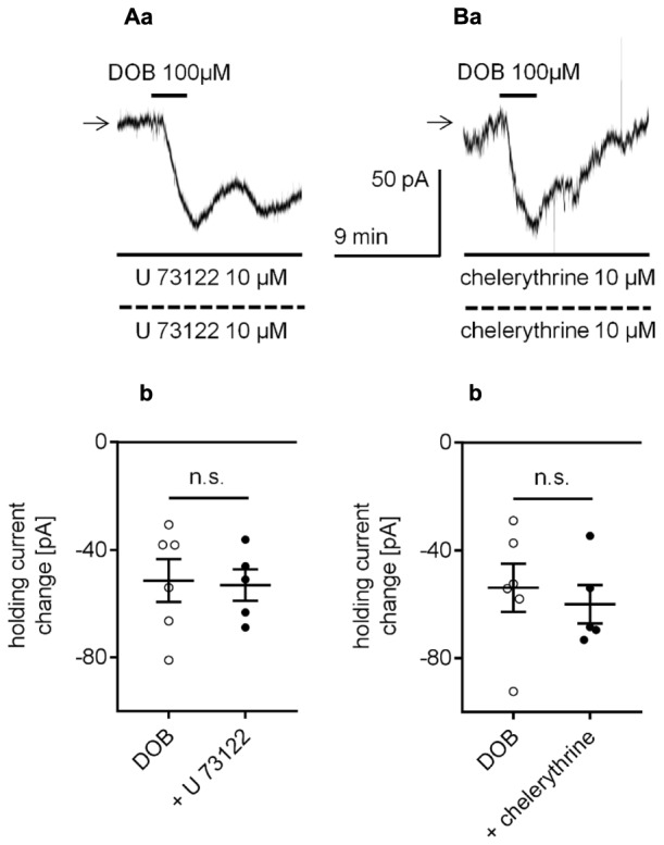
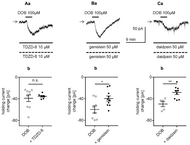
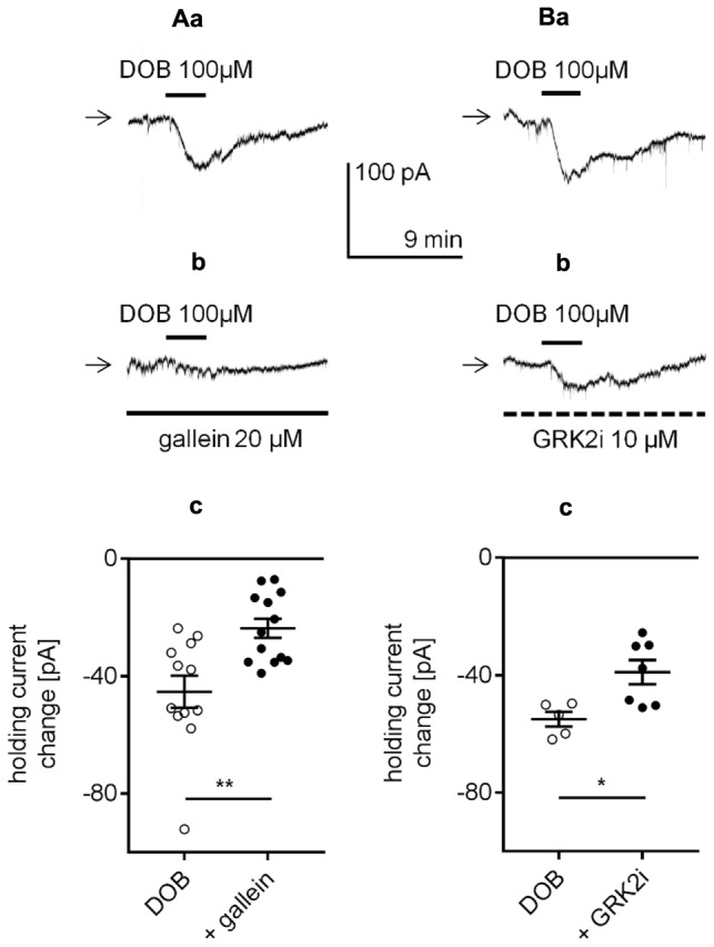
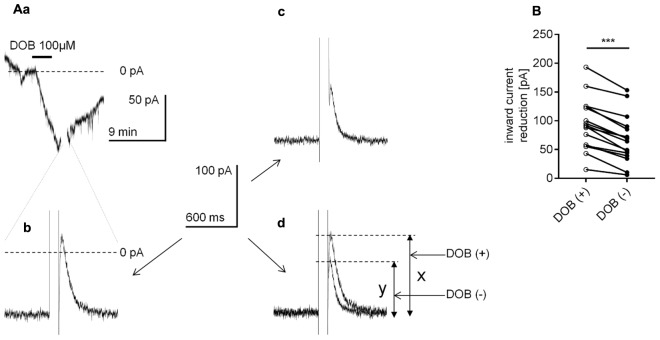
The medial prefrontal cortex (mPFC) receives dense noradrenergic projections from the locus coeruleus. Adrenergic innervation of mPFC pyramidal neurons plays an essential role in both physiology (control of memory formation, attention, working memory, and cognitive behavior) and pathophysiology (attention deficit hyperactivity disorder, posttraumatic stress disorder, cognitive deterioration after traumatic brain injury, behavioral changes related to addiction, Alzheimer's disease and depression). The aim of this study was to elucidate the mechanism responsible for adrenergic receptor-mediated control of the resting membrane potential in layer V mPFC pyramidal neurons. The membrane potential or holding current of synaptically isolated layer V mPFC pyramidal neurons was recorded in perforated-patch and classical whole-cell configurations in slices from young rats. Application of noradrenaline (NA), a neurotransmitter with affinity for all types of adrenergic receptors, evoked depolarization or inward current in the tested neurons irrespective of whether the recordings were performed in the perforated-patch or classical whole-cell configuration. The effect of noradrenaline depended on β1- and not α1- or α2-adrenergic receptor stimulation. Activation of β1-adrenergic receptors led to an increase in inward Na+ current through hyperpolarization-activated cyclic nucleotide-gated (HCN) channels, which carry a mixed Na+/K+ current. The protein kinase A- and C-, glycogen synthase kinase-3β- and tyrosine kinase-linked signaling pathways were not involved in the signal transduction between β1-adrenergic receptors and HCN channels. The transduction system operated in a membrane-delimited fashion and involved the βγ subunit of G-protein. Thus, noradrenaline controls the resting membrane potential and holding current in mPFC pyramidal neurons through β1-adrenergic receptors, which in turn activate HCN channels via a signaling pathway involving the βγ subunit.

Keywords: HCN channel; adrenergic receptors; holding current; membrane potential; prefrontal cortex; pyramidal neurons; rats; βγ subunit.


Figures

FIGURE 1
FIGURE 1
Effect of noradrenaline (NA) on the neuronal excitability of layer V medial prefrontal cortex (mPFC) pyramidal neurons in the absence of TTX. (A) IR-DIC image of a typical PFC slice with layer V pyramidal neurons (marked with white arrows). (B) Membrane potential depolarization evoked by bath application of NA (50 μM) recorded in the current-clamp classical whole-cell configuration. (C) Representative traces obtained from one neuron showing the response to a depolarizing current step (+150 pA, 1000 ms) before (control, a), during (NA 50 μM, b) and after NA bath application (washout, c). (D) Mean number of spikes evoked by depolarizing current steps (from +50 to +350 pA in 50 pA increments) before (control, open circles), during (NA 50 μM, black circles) and after (washout, gray circles) NA bath application. Horizontal arrows shown in this and other figures indicate the resting membrane potential or control holding current level. Continuous horizontal bars above the recording traces indicate the bath application of the agonists in this and other figures.
FIGURE 2
FIGURE 2
Effect of NA on the membrane potential and holding current in layer V mPFC pyramidal neurons. (A) Concentration-response curve for the effect of NA on the amplitude of the membrane potential depolarization. (B) Membrane potential depolarization evoked by bath application of NA (50 μM) recorded in the current-clamp perforated-patch (a) and classical whole-cell (b) configurations. Inward current evoked by bath application of NA (50 μM) recorded in the voltage-clamp perforated-patch (c) and classical whole-cell (d) configurations. (C) Depolarization amplitudes evoked by NA (50 μM) recorded in the current-clamp perforated-patch and classical whole-cell configurations (a). Inward current amplitudes evoked by NA (50 μM) recorded in the voltage-clamp perforated-patch and classical whole-cell configurations (b); ∗∗∗p < 0.001; n.s., non-significant. In this (Ca,b) and other figures, amplitudes of membrane potentials are shown as M ± SE and the distribution of individual measurements.
FIGURE 3
FIGURE 3
Effect of α1-adrenergic receptor stimulation on the membrane potential of layer V mPFC pyramidal neurons. (A) Membrane potential recorded during bath application of phenylephrine (100 μM, a). Amplitude of the membrane potential change evoked by 100 μM phenylephrine (phenylephrine, b). (B) Membrane potential change evoked by bath application of cirazoline (100 μM, a). Amplitude of the membrane potential change evoked by 100 μM cirazoline (cirazoline, b). (C) Membrane potential change evoked by bath application of cirazoline (100 μM) in the presence of prazosin (100 μM in the bath, a). Amplitude of the membrane potential change evoked by 100 μM cirazoline alone (cirazoline) and 100 μM cirazoline in the presence of 100 μM prazosin (+ prazosin, b). (D) Membrane potential change evoked by bath application of cirazoline (100 μM) in the presence of efaroxan (100 μM in the bath, a). Amplitude of the membrane potential change evoked by 100 μM cirazoline alone (cirazoline) and 100 μM cirazoline in the presence of 100 μM efaroxan (+ efaroxan, b); ∗∗∗p < 0.001; n.s., non-significant. Continuous horizontal bars below the recording traces indicate the bath/extracellular solution presence of the compounds in this and other figures.
FIGURE 4
FIGURE 4
Effect of α2-adrenergic receptor stimulation on the membrane potential of layer V mPFC pyramidal neurons. (A) Membrane potential recorded during bath application of medetomidine (100 μM, a). Amplitude of the membrane potential change evoked by 100 μM medetomidine (medetomidine, b). (B) Membrane potential change evoked by bath application of clonidine (100 μM, a). Amplitude of the membrane potential change evoked by 100 μM clonidine (clonidine, b). (C) Membrane potential change evoked by bath application of clonidine (100 μM) in the presence of yohimbine (60 μM in the bath, a). Amplitude of the membrane potential change evoked by 100 μM clonidine alone (clonidine) and 100 μM clonidine in the presence of 60 μM yohimbine (+ yohimbine, b). (D) Membrane potential changes evoked by bath application of ZD 7288 (50 μM) alone (a) and bath application of clonidine (100 μM) in the presence of ZD 7288 (50 μM in the bath, b). Amplitude of the membrane potential change evoked by 100 μM clonidine alone (clonidine) and 100 μM clonidine in the presence of 50 μM ZD 7288 (+ ZD 7288, c); ∗∗∗p < 0.001; n.s., non-significant.
FIGURE 5
FIGURE 5
Effect of selective β-adrenergic receptor blockers on NA-induced depolarization (A,B) and the inward current (C,D) in layer V mPFC pyramidal neurons. (A) Depolarization evoked by bath application of NA (50 μM) in the presence of the selective β1-adrenergic receptor blocker metoprolol (60 μM in the bath, a). Amplitude of the membrane potential change evoked by 50 μM NA alone (NA) and 50 μM NA in the presence of 60 μM metoprolol (+ metoprolol, b). (B) Depolarization evoked by bath application of NA (50 μM) in the presence of the selective β2-adrenergic receptor blocker ICI 118551 (50 μM in the bath, a). Amplitude of the membrane potential change evoked by 50 μM NA alone (NA) and 50 μM NA in the presence of 50 μM ICI 118551 (+ ICI 118551, b). (C) Inward current evoked by bath application of NA (50 μM) in the presence of the selective β1-adrenergic receptor blocker metoprolol (60 μM in the bath, a). Amplitude of the holding current change evoked by 50 μM NA alone (NA) and 50 μM NA in the presence of 60 μM metoprolol (+ metoprolol, b). (D) Inward current evoked by bath application of NA (50 μM) in the presence of the selective β2-adrenergic receptor blocker ICI 118551 (50 μM in the bath, a). Amplitude of the holding current change evoked by 50 μM NA alone (NA) and 50 μM NA in the presence of 50 μM ICI 118551 (+ ICI 118551, b); ∗∗p < 0.01; n.s., non-significant.
FIGURE 6
FIGURE 6
Effect of β1-adrenergic receptor stimulation on the holding current in layer V mPFC pyramidal neurons. (A) Inward currents evoked by bath application of the non-selective β-adrenergic receptor agonist isoproterenol alone (ISO, 100 μM, a) and isoproterenol (ISO, 100 μM) in the presence of the selective β1-adrenergic receptor blocker metoprolol (60 μM in the bath, b). Amplitude of the holding current change evoked by 100 μM isoproterenol alone (ISO) and 100 μM isoproterenol in the presence of 60 μM metoprolol (+ metoprolol, c). (B) Inward currents evoked by bath application of the selective β1-adrenergic receptor agonist dobutamine alone (DOB, 100 μM, a) and dobutamine (DOB, 100 μM) in the presence of the selective β1-adrenergic receptor blocker metoprolol (60 μM in the bath, b). Amplitude of the holding current change evoked by 100 μM dobutamine alone (DOB) and 100 μM dobutamine in the presence of 60 μM metoprolol (+ metoprolol, c); ∗∗∗p < 0.001. (C) Immunofluorescent staining of β1-adrenergic receptor protein in the rat mPFC. The signal is localized to neurons in different cortical layers (a). Layer V at higher magnification with a pyramidal neuron showing immunofluorescent signal within its soma (arrows) and apical dendrites (b). Scale bars (a) 100 μm, (b) 25 μm. M, medial; L, lateral; D, dorsal; V, ventral.
FIGURE 7
FIGURE 7
Identification of the cellular effector responsible for the β1-adrenergic receptor-dependent inward current in layer V mPFC pyramidal neurons. (A) Inward currents evoked by bath application of NA alone (50 μM, a) and by NA (50 μM) in the presence of a reduced Na+ concentration in the extracellular solution (b). Amplitude of the holding current change evoked by 50 μM NA alone (NA) and 50 μM NA in the presence of a reduced extracellular Na+ concentration (+ reduced Na+, c). (B) Inward currents evoked by bath application of NA alone (50 μM, a) and by NA (50 μM) in the presence of Cs+ (3 mM in the bath, b). Amplitude of the holding current change evoked by 50 μM NA alone (NA) and 50 μM NA in the presence of 3 mM Cs+ (+ Cs+, c). (C) Inward currents evoked by bath application of NA alone (50 μM, a) and by NA (50 μM) in the presence of ZD 7288 (50 μM in the bath, b). Amplitude of the holding current change evoked by 50 μM NA alone (NA) and 50 μM NA in the presence of 50 μM ZD 7288 (+ ZD 7288, c); ∗∗p < 0.01. (D) The membrane potential change (b) evoked by rectangular negative current steps (-400 pA, lasting 1000 ms, a). Amplified voltage sag shown in the inset. Histogram of the voltage sag size [%] in layer V mPFC pyramidal neurons (c). The size of the voltage sag was calculated by the difference between the maximum amplitude and sustained current response (x in Db) as a percentage of the maximum current response (y in Db).
FIGURE 8
FIGURE 8
Effect of the adenylyl cyclase (AC) and protein kinase A (PKA) inhibitors on the β1-adrenergic receptor-dependent inward current in layer V mPFC pyramidal neurons. (A) Inward current evoked by bath application of dobutamine (DOB, 100 μM) in the presence of the AC inhibitor (MDL 12330A, 20 μM in the bath a). Amplitude of the holding current change evoked by 100 μM dobutamine alone (DOB) and 100 μM dobutamine in the presence of 20 μM MDL 12330A (+ MDL 12330A, b). (B) Inward current evoked by bath application of dobutamine (DOB, 100 μM) in the presence of the AC inhibitor (SQ 22536, 100 μM in the bath, 1 mM in the pipette, a). Amplitude of the holding current change evoked by 100 μM dobutamine alone (DOB) and 100 μM dobutamine in the presence of 100 μM SQ 22536 in the bath and 1 mM in the pipette (+ SQ 22536, b). (C) Inward current evoked by bath application of dobutamine (DOB, 100 μM) in the presence of the PKA inhibitor (H 89, 10 μM in the bath and 10 μM in the pipette, a). Amplitude of the holding current change evoked by 100 μM dobutamine alone (DOB) and 100 μM dobutamine in the presence of 10 μM H 89 in the bath and 10 μM in the pipette (+ H 89, b); n.s., non-significant. Broken horizontal bars below the recording traces indicate the pipette/intracellular solution presence of the compounds in this and other figures.
FIGURE 9
FIGURE 9
Effect of the phospholipase C (PLC) and protein kinase C (PKC) inhibitors on the β1-adrenergic receptor-dependent inward current in layer V mPFC pyramidal neurons. (A) Inward current evoked by bath application of dobutamine (DOB, 100 μM) in the presence of the PLC inhibitor (U 73122, 10 μM in the bath and 10 μM in the pipette, a). Amplitude of the holding current change evoked by 100 μM dobutamine alone (DOB) and 100 μM dobutamine in the presence of 10 μM U 73122 in the bath and 10 μM in the pipette (+ U 73122, b). (B) Inward current evoked by bath application of dobutamine (DOB, 100 μM) in the presence of the PKC inhibitor (chelerythrine, 10 μM in the bath and 10 μM in the pipette, a). Amplitude of the holding current change evoked by 100 μM dobutamine alone (DOB) and 100 μM dobutamine in the presence of 10 μM chelerythrine in the bath and 10 μM in the pipette (+ chelerythrine, b); n.s., non-significant.
FIGURE 10
FIGURE 10
Effects of glycogen synthase kinase-3β (GSK-3β) and tyrosine kinase inhibitors on the β1-adrenergic receptor-dependent inward current in layer V mPFC pyramidal neurons. (A) Inward current evoked by bath application of dobutamine (DOB, 100 μM) in the presence of the GSK-3β inhibitor (TDZD-8, 10 μM in the bath and 10 μM in the pipette, a). Amplitude of the holding current change evoked by 100 μM dobutamine alone (DOB) and 100 μM dobutamine in the presence of 10 μM TDZD-8 in the bath and 10 μM in the pipette (+ TDZD-8, b). (B) Inward current evoked by bath application of dobutamine (DOB, 100 μM) in the presence of the tyrosine kinase inhibitor (genistein, 50 μM in the bath and 50 μM in the pipette, a). Amplitude of the holding current change evoked by 100 μM dobutamine alone (DOB) and 100 μM dobutamine in the presence of 50 μM genistein in the bath and 50 μM in the pipette (+ genistein, b). (C) Inward current evoked by bath application of dobutamine (DOB, 100 μM) in the presence of an inactive analog of genistein (daidzein, 50 μM in the bath and 50 μM in the pipette, a). Amplitude of the holding current change evoked by 100 μM dobutamine alone (DOB) and 100 μM dobutamine in the presence of 50 μM daidzein in the bath and 50 μM in the pipette (+ daidzein, b); p < 0.05; ∗∗p < 0.01; n.s., non-significant.
FIGURE 11
FIGURE 11
Effect of βγ subunit-dependent signaling inhibitors on the β1-adrenergic receptor-dependent inward current in layer V mPFC pyramidal neurons. (A) Inward currents evoked by bath application of dobutamine alone (DOB, 100 μM, a) and dobutamine (DOB, 100 μM) in the presence of gallein (20 μM in the bath, b). Amplitude of the holding current change evoked by 100 μM dobutamine alone (DOB) and 100 μM dobutamine in the presence of 20 μM gallein (+ gallein, c). The slices were also exposed to gallein in the extracellular solution for 2 h before current recordings. (B) Inward currents evoked by bath application of dobutamine alone (DOB, 100 μM, a) and dobutamine (DOB, 100 μM) in the presence of GRK2i (10 μM in the pipette solution, b). Amplitude of the holding current change evoked by 100 μM dobutamine in the absence (DOB) and presence of GRK2i 10 μM (+ GRK2i) (c). The currents were recorded >50 min after obtaining access to the cell; p < 0.05, ∗∗p < 0.01.
FIGURE 12
FIGURE 12
Effect of the voltage step on the β1-adrenergic receptor-dependent inward current in layer V mPFC pyramidal neurons. (A) Inward current evoked by bath application of dobutamine alone (DOB, 100 μM, a). At the peak of the inward current, the recording was interrupted, and a 100-ms depolarizing voltage step from the holding potential to +80 mV was applied. Following the voltage step, the inward current was reduced (b). At 20 min after termination of dobutamine application, when the recorded holding current returned to its resting control level, a current was injected from the pipette to obtain the maximum inward current level similar to that during the application of dobutamine. At this current level, a 100-ms depolarizing voltage step from the holding potential to +80 mV was applied again. Following the voltage step, the inward current was reduced (c). In (d), the overlapped current recordings from (b,c) are shown. The amplitude of the current reduction in the presence (x) and absence (y) of dobutamine is indicated. (B) Amplitude of the inward current reduction after the voltage step during dobutamine application [DOB (+)] and in the absence of dobutamine [DOB (–)] in the extracellular solution; ∗∗∗p < 0.001.

Similar articles

Cited by

References

    1. Agster K. L., Mejias-Aponte C. A., Clark B. D., Waterhouse B. D. (2013). Evidence for a regional specificity in the density and distribution of noradrenergic varicosities in rat cortex. J. Comp. Neurol. 521 2195–2207. 10.1002/cne.23270 - DOI - PMC - PubMed
    1. Akaike N. (1999). Gramicidin perforated patch recording technique. Folia Pharmacol. Japon. 113 339–347. 10.1254/fpj.113.339 - DOI - PubMed
    1. Akaike N., Harata N. (1994). Nystatin perforated patch recording and its applications to analyses of intracellular mechanisms. Jpn. J. Physiol. 44 433–473. 10.2170/jjphysiol.44.433 - DOI - PubMed
    1. Albarrán-Juárez J., Gilsbach R., Piekorz R. P., Pexa K., Beetz N., Schneider J., et al. (2009). Modulation of α2-adrenoceptor functions by heterotrimeric gαi protein isoforms. J. Pharmacol. Exp. Ther. 331 35–44. 10.1124/jpet.109.157230 - DOI - PMC - PubMed
    1. Andrews G. D., Lavin A. (2006). Methylphenidate increases cortical excitability via activation of alpha-2 noradrenergic receptors. Neuropsychopharmacology 31 594–601. 10.1038/sj.npp.1300818 - DOI - PMC - PubMed

LinkOut - more resources