

Lipid Uptake, Metabolism, and Transport in the Larval Zebrafish
- PMID: 29209275
- PMCID: PMC5701920
- DOI: 10.3389/fendo.2017.00319
Lipid Uptake, Metabolism, and Transport in the Larval Zebrafish
Abstract
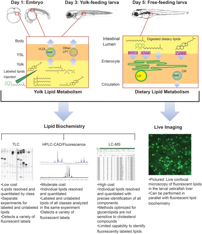
The developing zebrafish is a well-established model system for studies of energy metabolism, and is amenable to genetic, physiological, and biochemical approaches. For the first 5 days of life, nutrients are absorbed from its endogenous maternally deposited yolk. At 5 days post-fertilization, the yolk is exhausted and the larva has a functional digestive system including intestine, liver, gallbladder, pancreas, and intestinal microbiota. The transparency of the larval zebrafish, and the genetic and physiological similarity of its digestive system to that of mammals make it a promising system in which to address questions of energy homeostasis relevant to human health. For example, apolipoprotein expression and function is similar in zebrafish and mammals, and transgenic animals may be used to examine both the transport of lipid from yolk to body in the embryo, and the trafficking of dietary lipids in the larva. Additionally, despite the identification of many fatty acid and lipid transport proteins expressed by vertebrates, the cell biological processes that mediate the transport of dietary lipids from the intestinal lumen to the interior of enterocytes remain to be elucidated. Genetic tractability and amenability to live imaging and a range of biochemical methods make the larval zebrafish an ideal model in which to address open questions in the field of lipid transport, energy homeostasis, and nutrient metabolism.
Keywords: comparative physiology; enterocytes; lipid metabolism; lipoproteins; zebrafish.
Figures
Similar articles
-
Microsomal triglyceride transfer protein is required for yolk lipid utilization and absorption of dietary lipids in zebrafish larvae.Biochemistry. 2006 Dec 26;45(51):15179-87. doi: 10.1021/bi0619268. Epub 2006 Dec 1. Biochemistry. 2006. PMID: 17176039
-
Dietary cholesterol and apolipoprotein A-I are trafficked in endosomes and lysosomes in the live zebrafish intestine.Am J Physiol Gastrointest Liver Physiol. 2019 Mar 1;316(3):G350-G365. doi: 10.1152/ajpgi.00080.2018. Epub 2019 Jan 10. Am J Physiol Gastrointest Liver Physiol. 2019. PMID: 30629468 Free PMC article.
-
Using fluorescent lipids in live zebrafish larvae: From imaging whole animal physiology to subcellular lipid trafficking.Methods Cell Biol. 2016;133:165-78. doi: 10.1016/bs.mcb.2016.04.011. Epub 2016 May 9. Methods Cell Biol. 2016. PMID: 27263413 Free PMC article.
-
Zebrafish: gaining popularity in lipid research.Biochem J. 2010 Jul 15;429(2):235-42. doi: 10.1042/BJ20100293. Biochem J. 2010. PMID: 20578994 Review.
-
Zebrafish ApoB-Containing Lipoprotein Metabolism: A Closer Look.Arterioscler Thromb Vasc Biol. 2024 May;44(5):1053-1064. doi: 10.1161/ATVBAHA.123.318287. Epub 2024 Mar 14. Arterioscler Thromb Vasc Biol. 2024. PMID: 38482694 Free PMC article. Review.
Cited by
-
Use of Zebrafish in Drug Discovery Toxicology.Chem Res Toxicol. 2020 Jan 21;33(1):95-118. doi: 10.1021/acs.chemrestox.9b00335. Epub 2019 Nov 16. Chem Res Toxicol. 2020. PMID: 31625720 Free PMC article. Review.
-
Sustainable aquafeed development: Incorporating select fruit wastes into Zebrafish diets using mathematical model-based approach.Saudi J Biol Sci. 2023 Nov;30(11):103834. doi: 10.1016/j.sjbs.2023.103834. Epub 2023 Oct 12. Saudi J Biol Sci. 2023. PMID: 37885611 Free PMC article.
-
Mass Spectrometry-Based Zebrafish Toxicometabolomics: A Review of Analytical and Data Quality Challenges.Metabolites. 2021 Sep 17;11(9):635. doi: 10.3390/metabo11090635. Metabolites. 2021. PMID: 34564451 Free PMC article. Review.
-
Deficiency in the autophagy modulator Dram1 exacerbates pyroptotic cell death of Mycobacteria-infected macrophages.Cell Death Dis. 2020 Apr 24;11(4):277. doi: 10.1038/s41419-020-2477-1. Cell Death Dis. 2020. PMID: 32332700 Free PMC article.
-
Deletion of the chd7 Hinders Oligodendrocyte Progenitor Cell Development and Myelination in Zebrafish.Int J Mol Sci. 2023 Aug 31;24(17):13535. doi: 10.3390/ijms241713535. Int J Mol Sci. 2023. PMID: 37686337 Free PMC article.
References
Publication types
Grants and funding
LinkOut - more resources
Full Text Sources
Other Literature Sources
Molecular Biology Databases