. 2017:2017:2945012.
doi: 10.1155/2017/2945012. Epub 2017 Oct 25.

Functional Characterization of Preadipocytes Derived from Human Periaortic Adipose Tissue

Affiliations

Functional Characterization of Preadipocytes Derived from Human Periaortic Adipose Tissue

Diana Vargas et al. Int J Endocrinol. 2017.

Abstract

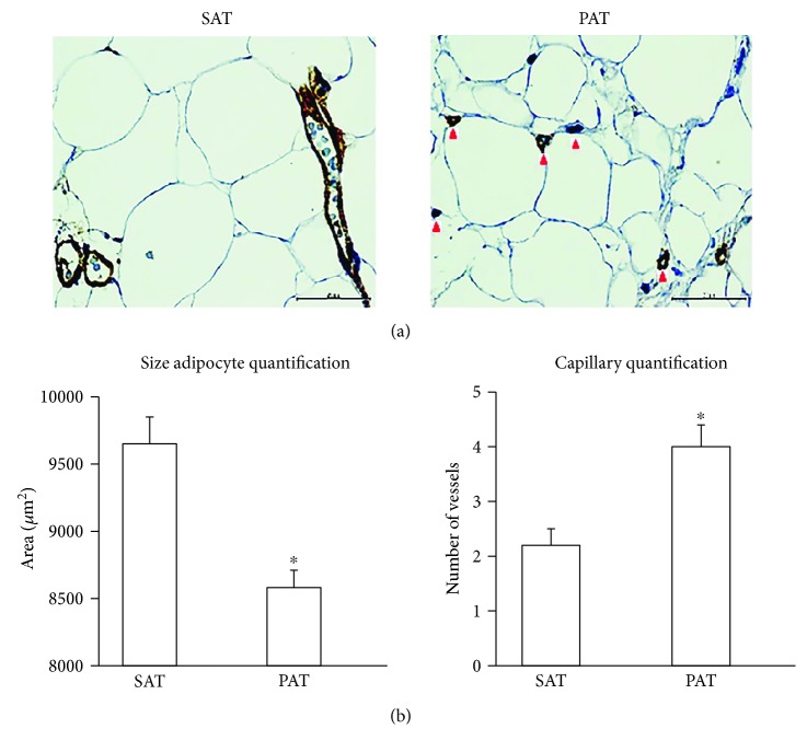
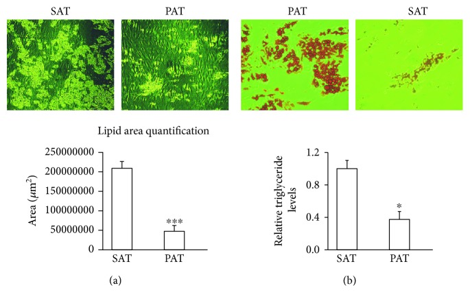
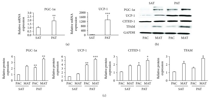
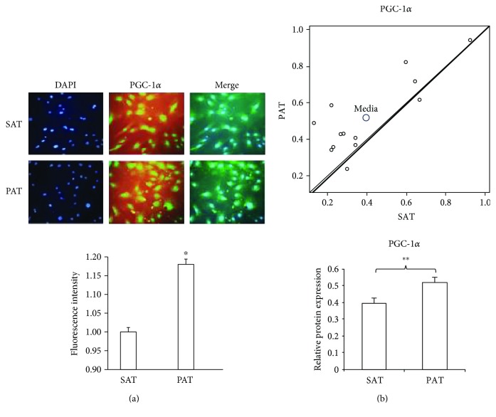
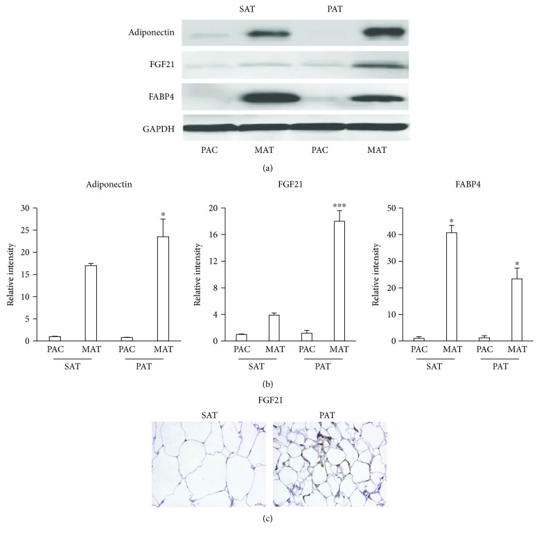
Adipose tissue can affect the metabolic control of the cardiovascular system, and its anatomic location can affect the vascular function differently. In this study, biochemical and phenotypical characteristics of adipose tissue from periaortic fat were evaluated. Periaortic and subcutaneous adipose tissues were obtained from areas surrounding the ascending aorta and sternotomy incision, respectively. Adipose tissues were collected from patients undergoing myocardial revascularization or mitral valve replacement surgery. Morphological studies with hematoxylin/eosin and immunohistochemical assay were performed in situ to quantify adipokine expression. To analyze adipogenic capacity, adipokine expression, and the levels of thermogenic proteins, adipocyte precursor cells were isolated from periaortic and subcutaneous adipose tissues and induced to differentiation. The precursors of adipocytes from the periaortic tissue accumulated less triglycerides than those from the subcutaneous tissue after differentiation and were smaller than those from subcutaneous adipose tissue. The levels of proteins involved in thermogenesis and energy expenditure increased significantly in periaortic adipose tissue. Additionally, the expression levels of adipokines that affect carbohydrate metabolism, such as FGF21, increased significantly in mature adipocytes induced from periaortic adipose tissue. These results demonstrate that precursors of periaortic adipose tissue in humans may affect cardiovascular events and might serve as a target for preventing vascular diseases.


Figures

Figure 1
Figure 1
Histological features and the size of adipocytes from PAT and SAT. (a) PAT and SAT were fixed in 10% formaldehyde and used for morphological analysis and the determination of adipocyte size and capillary number. Immunohistochemistry revealed brown intracytoplasmic precipitates only in the intima marked by von Willebrand factor staining (red arrow). (b) Adipocyte size was determined by analyzing five quadrants in each image by ImageJ software. The capillary number was determined by counting 15 random fields in three different plates at a magnification of 40x. p < 0.05 indicates a statistically significant difference in the size of adipocytes from PAT versus SAT.
Figure 2
Figure 2
Adipogenic capacity from precursors of PAT and SAT. (a) Precursor cells from PAT and SAT adipocytes were induced to differentiate into mature adipocytes. Images were acquired after 15 days of differentiation. (b) The surface area of accumulated lipids was determined by dividing the image into four quadrants, which were further subdivided into five zones per quadrant. Data are expressed as means ± SD (n = 4). p < 0.05 indicates the differences in the accumulation of triglycerides between SAT and PAT. ∗∗∗p < 0.001 indicates a statistically significant difference in lipid accumulation in differentiated adipocytes from PAT versus SAT. Relative levels of triglycerides in SAT and PAT were evaluated after the cells reached differentiation. To quantify triglycerides, 1 mL of isopropanol was added for 5 min, to distain the fat deposits. Absorbance was measured at 510 mm wavelength.
Figure 3
Figure 3
PAT expressed proteins involved in thermogenesis. (a) Samples from SAT and PAT were obtained and immediately frozen in liquid nitrogen. mRNA was extracted and the detection of PGC-1α and UCP-1 was performed by qPCR. (b) Precursor adipose cells (PAC) from SAT and PAT were induced to differentiate into mature adipocytes (MAT), and proteins were extracted to quantify the levels of PGC-1α, UCP-1, CITED1, and TFAM by Western blotting. (c) Relative intensity of the protein bands was determined by densitometry. Data were normalized to the housekeeping protein GAPDH and expressed as means ± SD (n = 4). p < 0.05, in three different studies, indicates a statistically significant difference in protein levels in precursor adipose cells (SAT versus PAT) and mature adipocyte cells (SAT versus PAT). ∗∗p < 0.01 indicates the difference between PAT and SAT both in precursor adipocytes and mature adipocytes. ∗∗∗p < 0.01 indicates the difference between PAT and SAT in mature adipocytes.
Figure 4
Figure 4
PGC-1α expression and the relationship between PAT and SAT. (a) Adipocyte precursor cells were isolated from PAT and SAT, induced to proliferate, fixed in 10% formaldehyde, and stained for the presence of PGC-1α by immunofluorescence. The fluorescent intensity was quantified with ImageJ software. Data are expressed as means ± SD. p < 0.05 indicates the differences in the fluorescent intensity between SAT and PAT. Data correspond to the three different studies. (b) The scatter plot shows the relationship for the PGC-1α level in precursor cells from PAT and SAT. Each data point represents a patient. The y-axis (Y) shows the trend toward PAT expression, and the x-axis (X) shows the trend toward SAT expression (n = 13). The bars indicate intensity levels as determined by densitometry of the bands between SAT and PAT from each patient using paired Student's t-test of Western blot analysis of PGC-1α. ∗∗p < 0.01 indicates significative differences in PGC-1α protein expression between SAT and PAT.
Figure 5
Figure 5
Expression of adipokines in PAT. (a) Adipocyte precursor cells (PAC) from SAT and PAT were induced to differentiate into mature adipocytes (MAT). Proteins were extracted to quantify the levels of adiponectin, FGF21, and FABP4 by Western blotting. (b) Relative intensity of the protein bands was determined by densitometry. Data were normalized to the housekeeping protein GAPDH and expressed as means ± SD (n = 4). (c) Specimens were fixed in 10% formaldehyde and stained for the presence of FGF21 by immunohistochemistry. Brown precipitates were indicative of FGF21 within adipocytes. Data were normalized to the housekeeping gene GAPDH. p < 0.05 indicates differences between PAT mature cells in relation to SAT mature cells. ∗∗∗p < 0.01 indicates differences between PAT mature cells and SAT mature cells.

References

    1. Gil-Ortega M., Somoza B., Huang Y., Gollasch M., Fernandez-Alfonso M. S. Regional differences in perivascular adipose tissue impacting vascular homeostasis. Trends in Endocrinology & Metabolism. 2015;26(7):367–375. doi: 10.1016/j.tem.2015.04.003. - DOI - PubMed
    1. Houben A. J., Eringa E. C., Jonk A. M., Serne E. H., Smulders Y. M., Stehouwer C. D. Perivascular fat and the microcirculation: relevance to insulin resistance, diabetes, and cardiovascular disease. Current Cardiovascular Risk Reports. 2012;6(1):80–90. doi: 10.1007/s12170-011-0214-0. - DOI - PMC - PubMed
    1. Eringa E. C., Bakker W., van Hinsbergh V. W. Paracrine regulation of vascular tone, inflammation and insulin sensitivity by perivascular adipose tissue. Vascular Pharmacology. 2012;56(5-6):204–209. doi: 10.1016/j.vph.2012.02.003. - DOI - PubMed
    1. Lee R. M., Dickhout J. G., Sandow S. L. Vascular structural and functional changes: their association with causality in hypertension: models, remodeling and relevance. Hypertension Research. 2016;40(4):311–323. doi: 10.1038/hr.2016.145. - DOI - PubMed
    1. Sung H. K., Doh K. O., Son J. E., et al. Adipose vascular endothelial growth factor regulates metabolic homeostasis through angiogenesis. Cell Metabolism. 2013;17(1):61–72. doi: 10.1016/j.cmet.2012.12.010. - DOI - PubMed

LinkOut - more resources