

Guiding histological assessment of uterine lesions using 3D in vitro ultrasonography and stereotaxis
- PMID: 29209483
- PMCID: PMC5707776
Guiding histological assessment of uterine lesions using 3D in vitro ultrasonography and stereotaxis
Abstract
Objective: To compare ultrasonographic features of uterine lesions with the findings at macroscopy and microscopy.
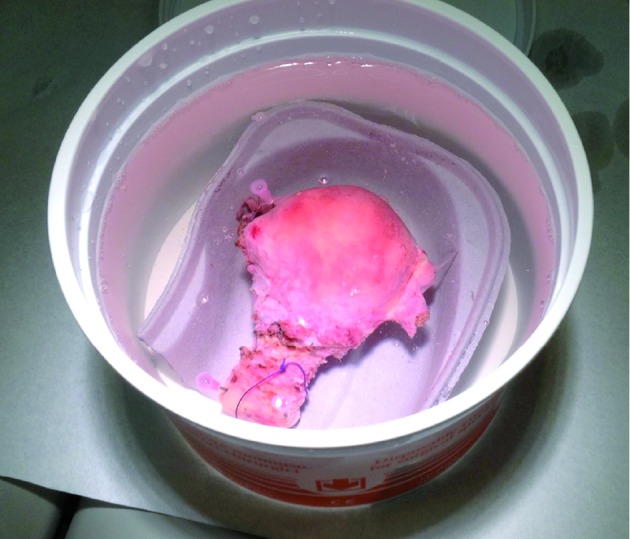
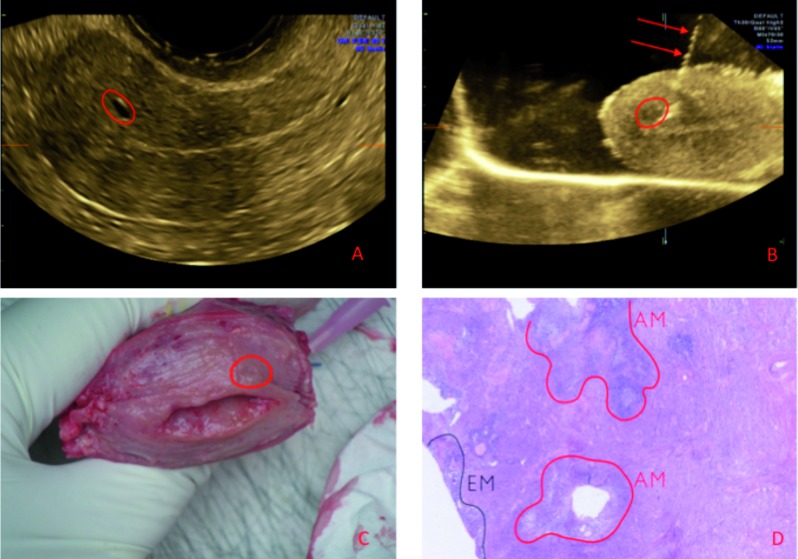
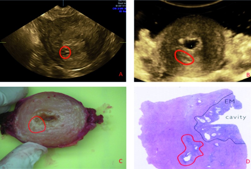
Methods: Case series of ten consecutive women undergoing a hysterectomy for uterine pathology. A preoperative transvaginal ultrasound examination was performed. After hysterectomy, the uterus was re-evaluated by 3D in vitro ultrasonography and in vitro gel instillation sonography (iGIS). The lesion of interest was pinpointed by inserting an intramuscular injection needle using a free-hand 2D-ultrasound guided technique to focus the macroscopic and the microscopic examination by the pathologist.
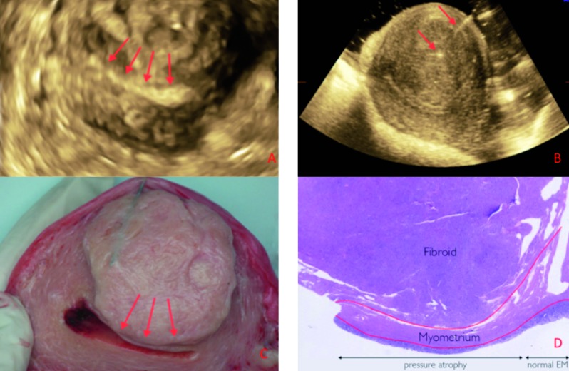
Results: Adenomyosis, benign fibroids and infiltrating endometrial cancer were diagnosed in six, five and one patient, respectively. We found that iGIS improved image quality of in vitro ultrasound. There was a good correlation between the reported ultrasound features and the final histological diagnosis. Some lesions had been misinterpreted during preoperative ultrasonography or at macroscopical examination: e.g. dense myometrial vessels reported as small myometrial cysts at grey scale ultrasound examination; absence of macroscopical lesions in a case of diffuse adenomyosis.
Conclusions: In vitro 3D ultrasonography and iGIS allow for accurate mapping of uterine lesions so that ultrasound features can be matched with final histology. Our series demonstrates some pitfalls in the interpretation of sonographic and macroscopic features of uterine lesions. Stereotaxis of focal uterine lesions could focus histological assessment and reduces examination time for the pathologist.
Keywords: 3D ultrasonography; adenomyosis; iGIS; in vitro; in vitro gel instillation sonography; myoma; stereotaxis.
Figures
Similar articles
-
Transvaginal sonographic features of diffuse adenomyosis in 18-30-year-old nulligravid women without endometriosis: association with symptoms.Ultrasound Obstet Gynecol. 2015 Dec;46(6):730-6. doi: 10.1002/uog.14834. Ultrasound Obstet Gynecol. 2015. PMID: 25728241
-
Three-dimensional ultrasound in diagnosis of adenomyosis: histologic correlation with ultrasound targeted biopsies of the uterus.J Minim Invasive Gynecol. 2013 Nov-Dec;20(6):803-10. doi: 10.1016/j.jmig.2013.05.002. J Minim Invasive Gynecol. 2013. PMID: 24183272
-
Optimizing the Histological Diagnosis of Adenomyosis Using in vitro Three-Dimensional Ultrasonography.Gynecol Obstet Invest. 2016;81(6):563-567. doi: 10.1159/000445072. Epub 2016 Mar 23. Gynecol Obstet Invest. 2016. PMID: 27002642
-
Diagnosing adenomyosis: an integrated clinical and imaging approach.Hum Reprod Update. 2020 Apr 15;26(3):392-411. doi: 10.1093/humupd/dmz049. Hum Reprod Update. 2020. PMID: 32097456 Review.
-
Ultrasound diagnosis of uterine myomas.Minerva Ginecol. 2016 Jun;68(3):297-312. Epub 2016 Mar 25. Minerva Ginecol. 2016. PMID: 27014801 Review.
Cited by
-
Recent advances in understanding and managing adenomyosis.F1000Res. 2019 Mar 13;8:F1000 Faculty Rev-283. doi: 10.12688/f1000research.17242.1. eCollection 2019. F1000Res. 2019. PMID: 30918629 Free PMC article. Review.
-
The Correlation of the IETA Ultrasound Score with the Histopathology Results for Women with Abnormal Bleeding in Western Romania.Diagnostics (Basel). 2021 Jul 26;11(8):1342. doi: 10.3390/diagnostics11081342. Diagnostics (Basel). 2021. PMID: 34441275 Free PMC article.
References
-
- Azziz R. Adenomyosis: current perspectives. Obstet Gynecol Clin North Am. 1989;16:221–235. - PubMed
-
- Bergholt T, Eriksen L, Berendt N. Prevalence and risk factors of adenomyosis at hysterectomy. Hum reprod. 2001;16:2418–2421. - PubMed
-
- Bird CC, McElin TW, Manalo-Estrella P. The elusive adenomyosis of the uterus revisited. Am J Obstet Gynecol. 1972;112:583–593. - PubMed
-
- Dueholm M. Transvaginal ultrasound for diagnosis of adenomyosis: a review. Best Pract Res Clin Obstet Gynaecol. 2006;20:569–582. - PubMed
-
- Fernandez H, Donnadieu AC. Adenomyosis. J Gynecol Obstet Biol reprod. 2007;36:179–185. - PubMed
LinkOut - more resources
Full Text Sources
Other Literature Sources