

A Case of Proliferative Glomerulonephritis with Monoclonal IgG Deposits That Showed Predominantly Membranous Features
- PMID: 29209544
- PMCID: PMC5676379
- DOI: 10.1155/2017/1027376
A Case of Proliferative Glomerulonephritis with Monoclonal IgG Deposits That Showed Predominantly Membranous Features
Abstract
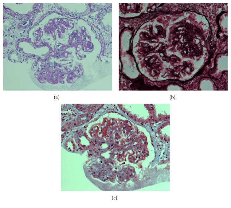
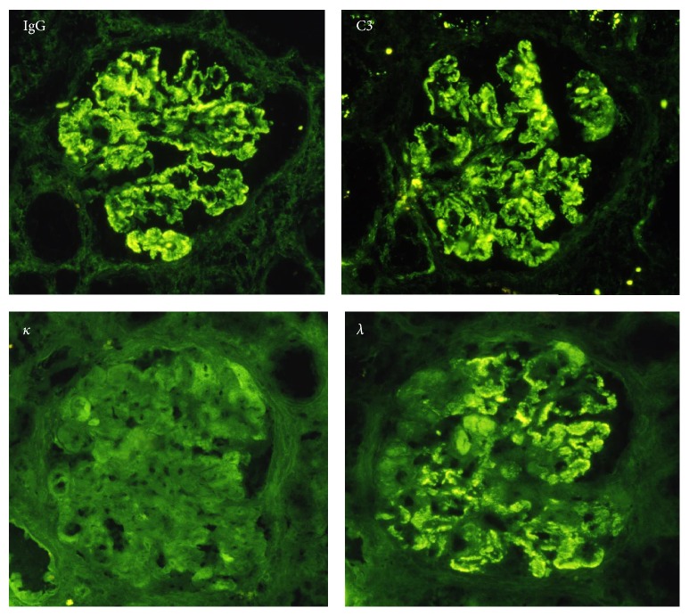
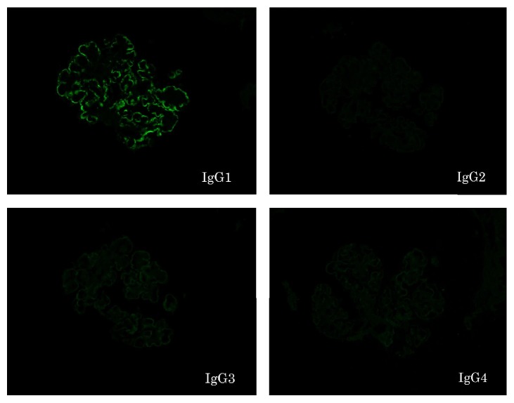
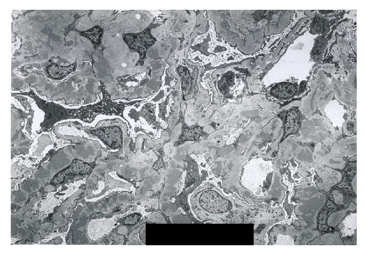
In 2004, the novel category of monoclonal IgG deposition disease has been proposed and termed "proliferative glomerulonephritis with monoclonal IgG deposits" (PGNMID). This disease is characterized by membranoproliferative glomerulonephritis and staining for a single light-chain isotype and gamma heavy-chain subclass. A 76-year-old male who had monoclonal gammopathy was referred to our hospital because of proteinuria. The renal biopsy showed diffuse thickening of the glomerular capillary walls with focal mesangial proliferation. On immunofluorescence study, only IgG1 among the four subclasses and lambda light chains were detected mainly in the glomerular capillary walls. From these results, we diagnosed our case as PGNMID showing predominantly membranous features. Almost all pathological findings on light microscopy of PGNMID are membranoproliferative GN or endocapillary proliferative GN, while membranous GN cases are rare. Here, we present the case of PGNMID that showed predominantly membranous features on light microscopy.
Figures
Similar articles
-
Discordance of light chain isotypes between serum and glomerular deposits in proliferative glomerulonephritis with monoclonal IgG deposits: a case report and review of the literature.BMC Nephrol. 2023 Jul 1;24(1):199. doi: 10.1186/s12882-023-03256-5. BMC Nephrol. 2023. PMID: 37393252 Free PMC article. Review.
-
Successful treatment with bortezomib and dexamethasone for proliferative glomerulonephritis with monoclonal IgG deposits in multiple myeloma: a case report.BMC Nephrol. 2017 Apr 6;18(1):127. doi: 10.1186/s12882-017-0524-7. BMC Nephrol. 2017. PMID: 28385149 Free PMC article.
-
Proliferative glomerulonephritis with monoclonal immunoglobulin deposits: a nephrologist perspective.Nephrol Dial Transplant. 2021 Jan 25;36(2):208-215. doi: 10.1093/ndt/gfz176. Nephrol Dial Transplant. 2021. PMID: 33494099 Review.
-
Chronological change of renal pathological findings in the proliferative glomerulonephritis with monoclonal IgG deposits considered to have recurred early after kidney transplantation.CEN Case Rep. 2019 Aug;8(3):151-158. doi: 10.1007/s13730-019-00384-6. Epub 2019 Feb 25. CEN Case Rep. 2019. PMID: 30805792 Free PMC article.
-
Proliferative glomerulonephritis with monoclonal IgG2κ deposit successfully treated with steroids: a case report and review of the literature.CEN Case Rep. 2013 Nov;2(2):197-203. doi: 10.1007/s13730-013-0064-3. Epub 2013 Feb 26. CEN Case Rep. 2013. PMID: 28509293 Free PMC article.
Cited by
-
Kidney Diseases: The Age of Molecular Markers.Adv Exp Med Biol. 2021;1306:13-27. doi: 10.1007/978-3-030-63908-2_2. Adv Exp Med Biol. 2021. PMID: 33959903
-
Hemato-Renal Profile of Proliferative Glomerulonephritis with Monoclonal Immunoglobulin Deposits.Indian J Nephrol. 2024 Jul 22;35(3):355-367. doi: 10.25259/ijn_489_23. Indian J Nephrol. 2024. PMID: 39850252 Free PMC article.
-
Concurrent presentation of proliferative glomerulonephritis with monoclonal immunoglobulin deposits and light chain proximal tubulopathy: a case report and review of the literature.Front Med (Lausanne). 2025 Jan 31;12:1502798. doi: 10.3389/fmed.2025.1502798. eCollection 2025. Front Med (Lausanne). 2025. PMID: 39958827 Free PMC article.
-
Discordance of light chain isotypes between serum and glomerular deposits in proliferative glomerulonephritis with monoclonal IgG deposits: a case report and review of the literature.BMC Nephrol. 2023 Jul 1;24(1):199. doi: 10.1186/s12882-023-03256-5. BMC Nephrol. 2023. PMID: 37393252 Free PMC article. Review.
References
-
- de Seigneux S., Bindi P., Debiec H., et al. Immunoglobulin Deposition Disease With a Membranous Pattern and a Circulating Monoclonal Immunoglobulin G With Charge-Dependent Aggregation Properties. American Journal of Kidney Diseases. 2010;56(1):117–121. doi: 10.1053/j.ajkd.2010.03.010. - DOI - PubMed
Publication types
LinkOut - more resources
Full Text Sources
Other Literature Sources
