Review
. 2017 Nov 20:5:244.
doi: 10.3389/fped.2017.00244. eCollection 2017.

Meckel-Gruber Syndrome: An Update on Diagnosis, Clinical Management, and Research Advances

Affiliations
Review

Meckel-Gruber Syndrome: An Update on Diagnosis, Clinical Management, and Research Advances

Verity Hartill et al. Front Pediatr. .

Abstract

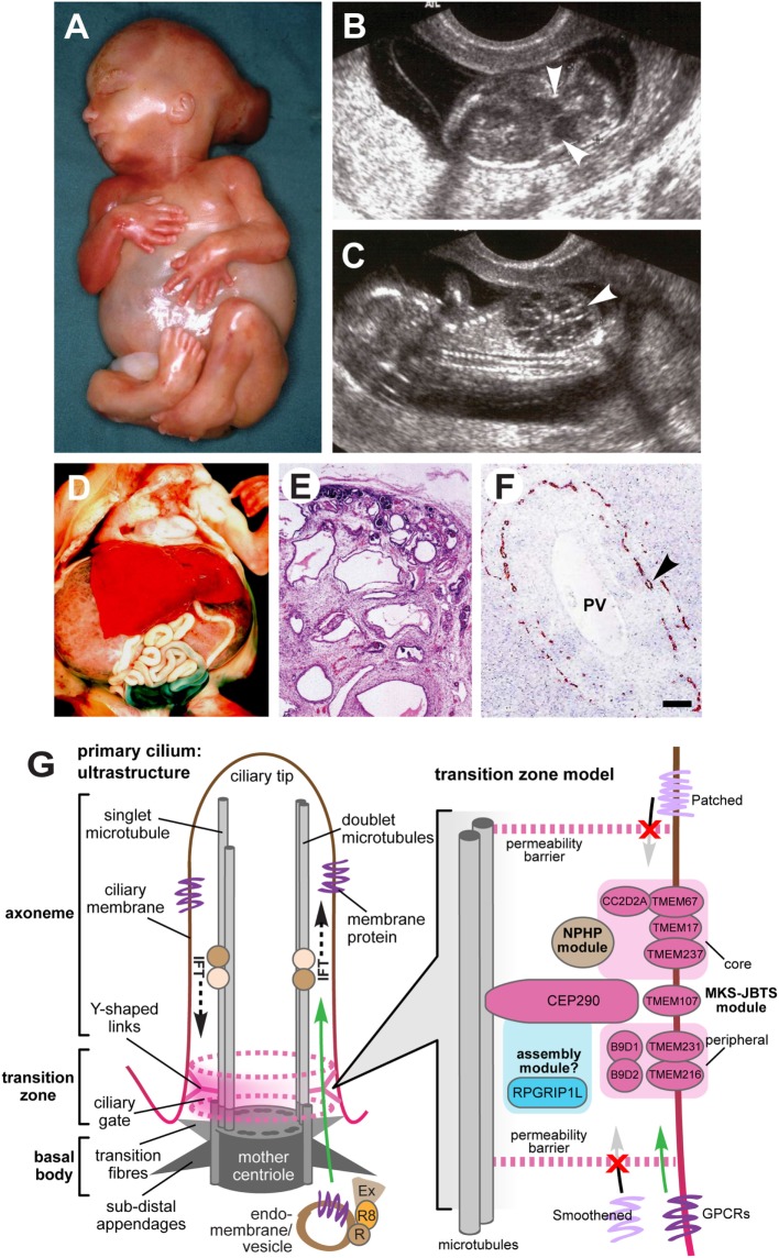
Meckel-Gruber syndrome (MKS) is a lethal autosomal recessive congenital anomaly syndrome caused by mutations in genes encoding proteins that are structural or functional components of the primary cilium. Conditions that are caused by mutations in ciliary genes are collectively termed the ciliopathies, and MKS represents the most severe condition in this group of disorders. The primary cilium is a microtubule-based organelle, projecting from the apical surface of vertebrate cells. It acts as an "antenna" that receives and transduces chemosensory and mechanosensory signals, but also regulates diverse signaling pathways, such as Wnt and Shh, that have important roles during embryonic development. Most MKS proteins localize to a distinct ciliary compartment called the transition zone (TZ) that regulates the trafficking of cargo proteins or lipids. In this review, we provide an up-to-date summary of MKS clinical features, molecular genetics, and clinical diagnosis. MKS has a highly variable phenotype, extreme genetic heterogeneity, and displays allelism with other related ciliopathies such as Joubert syndrome, presenting significant challenges to diagnosis. Recent advances in genetic technology, with the widespread use of multi-gene panels for molecular testing, have significantly improved diagnosis, genetic counseling, and the clinical management of MKS families. These include the description of some limited genotype-phenotype correlations. We discuss recent insights into the molecular basis of disease in MKS, since the functions of some of the relevant ciliary proteins have now been determined. A common molecular etiology appears to be disruption of ciliary TZ structure and function, affecting essential developmental signaling and the regulation of secondary messengers.

Keywords: Meckel–Gruber syndrome; Shh signaling; oligohydramnios; primary cilium; renal cystic dysplasia; transition zone.


Figures

Figure 1
Figure 1
Clinical features of Meckel–Gruber syndrome (MKS) and schematic of primary cilia structure. (A) Typical external features for a fetus with MKS at gestation age 16/40, showing typical clinical features comprising occipital encephalocele, massive flank masses due to bilateral renal cystic dysplasia, postaxial hexadactyly of all limbs, and a typical Potter’s sequence facies with a slanting forehead and flattened nose. (B,C) Ultrasound findings at 14+/40 weeks of gestation for MKS showing (B) encephalocele (arrowheads), and (C) large cystic kidneys (arrowhead). (D) Massive swelling of the abdomen of a fetus at gestation age 18+/40 with MKS due to grossly enlarged, cystic kidneys. (E) Hematoxylin-eosin staining of MKS fetal kidney at gestation age 18+/40 showing cystic dysplasia, comprising large, fluid-filled cysts, small cysts, and cystic swelling of the proximal tubules and glomeruli, with the absence of normal renal parenchyma. (F) Immunohistochemical staining of MKS fetal liver at gestation age 18+/40 for cytokeratin-19, showing the retention of embryonic bile duct structures (the ductal plate malformation) without the formation of patent bile ducts (arrowhead). PV, hepatic portal vein. (G) Left: simplified schematic of cilium ultrastructure (individual components are not to scale). For the purposes of clarity, the ciliary axoneme is represented by two doublets of microtubules (the A- and B-tubules; gray rods), and the nine-fold symmetry in the structure is indicated by dark gray ovals in the mother centriole. The axoneme is bound by ciliary membrane (red/brown line and shading). The ciliary transition zone (TZ) is characterized by Y-shaped links (pink) that mediate interactions with the ciliary membrane. The permeability barrier called the “ciliary gate” is indicated by the dashed pink ovals and pink shading, and is thought to consist of transition fibers and TZ proteins. Ciliary cargo proteins (purple) are trafficking by vesicular transport mediated by coordinated interactions between the Rab family of small GTPases (RAB11 and RAB8; R8), the RAB8 activator protein Rabin8 (R), and the exocyst protein complex (Ex) at the ciliary base. The exocyst tethers vesicles to the periciliary membrane, facilitating transport through the ciliary gate (green arrow), followed by transport within the cilium mediated by intraflagellar transport (IFT; dashed black arrows). Right: schematic of a TZ model, showing the possible location of TZ proteins mutated in MKS and related ciliopathies (pink ovals) forming the “MKS-JBTS module,” RPGRIP1L (blue oval) which is thought to be a component of a ciliary assembly module, and a third functional module (brown) that is associated with nephronophthisis (NPHP) proteins. The selective transport of cargo transmembrane proteins (lilac or purple) is indicated by arrows. The proteins include G-protein-coupled receptors (GPCRs), and effectors of the Shh signaling pathway (Smoothened and Patched). Images (A–C) are used by kind permission of Dr. Riitta Salonen (Rinnekoti Foundation, Helsinki, Finland) from the Robert J. Gorlin Slide Collection. JBTS, Joubert syndrome.

References

    1. Meckel J. Beschreibung zweier, durch sehr ähnliche Bildungsabweichungen entstellter Geschwister. Dtsch Arch Physiol (1882) 7:99–172.
    1. Gruber G. Beitrage zur frage “gekoppelter” Miszbildungen (Akrocephalo-syndactylie und dysencephalia splanchnocystica). Beitr Pathol Anat (1934) 93:459–76.
    1. Opitz JM, Howe JJ. The Meckel syndrome (dysencephalia splanchnocystica, the Grüber syndrome). Birth Defects Orig Art Ser (1969) 2:167–79.
    1. Mecke S, Passarge E. Encephalocele, polycystic kidneys and polydactyly as an autosomal recessive trait simulating certain other disorders – Meckel syndrome. Ann Genet (1971) 14:97–103. - PubMed
    1. Fraser FC, Lytwyn A. Spectrum of anomalies in the Meckel syndrome, or maybe there is a malformation syndrome with at least one constant anomaly. Am J Med Genet (1981) 9:67–73.10.1002/ajmg.1320090112 - DOI - PubMed