. 2017 Nov 20:5:66.
doi: 10.3389/fbioe.2017.00066. eCollection 2017.

Culture Medium Supplements Derived from Human Platelet and Plasma: Cell Commitment and Proliferation Support

Affiliations

Culture Medium Supplements Derived from Human Platelet and Plasma: Cell Commitment and Proliferation Support

Anita Muraglia et al. Front Bioeng Biotechnol. .

Abstract

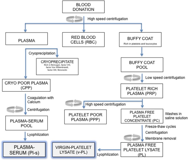
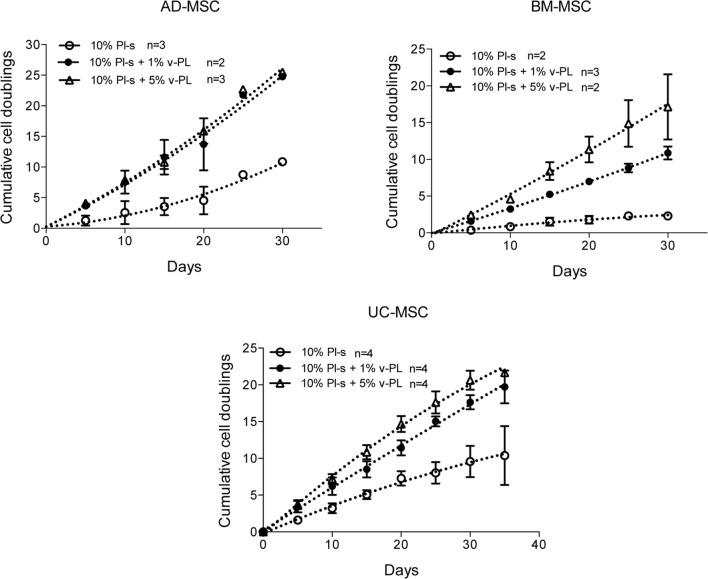
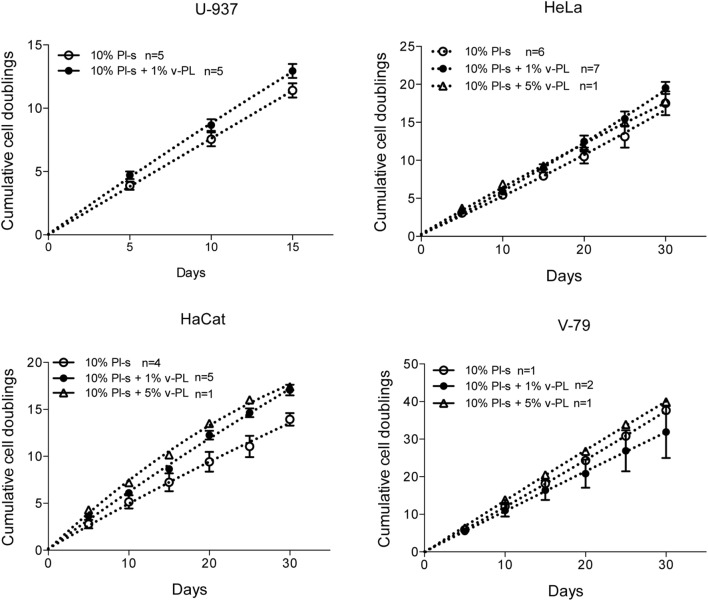
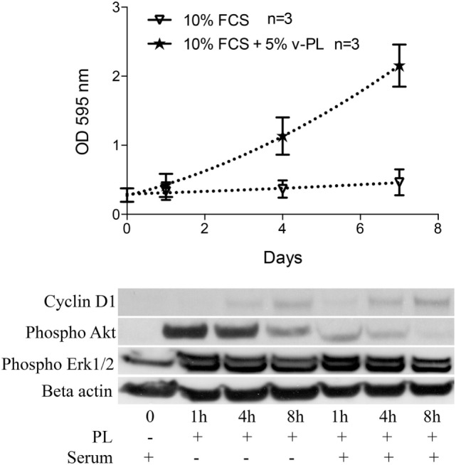
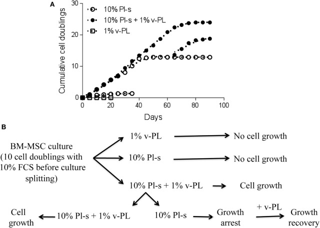
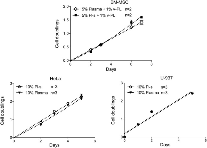
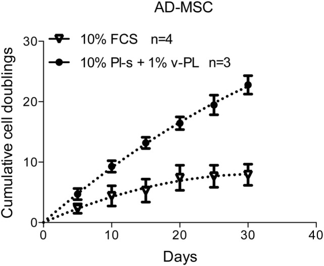
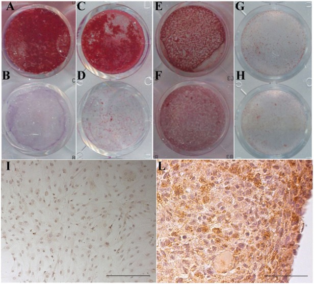
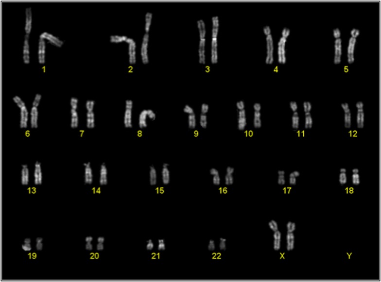
Present cell culture medium supplements, in most cases based on animal sera, are not fully satisfactory especially for the in vitro expansion of cells intended for human cell therapy. This paper refers to (i) an heparin-free human platelet lysate (PL) devoid of serum or plasma components (v-PL) and (ii) an heparin-free human serum derived from plasma devoid of PL components (Pl-s) and to their use as single components or in combination in primary or cell line cultures. Human mesenchymal stem cells (MSC) primary cultures were obtained from adipose tissue, bone marrow, and umbilical cord. Human chondrocytes were obtained from articular cartilage biopsies. In general, MSC expanded in the presence of Pl-s alone showed a low or no proliferation in comparison to cells grown with the combination of Pl-s and v-PL. Confluent, growth-arrested cells, either human MSC or human articular chondrocytes, treated with v-PL resumed proliferation, whereas control cultures, not supplemented with v-PL, remained quiescent and did not proliferate. Interestingly, signal transduction pathways distinctive of proliferation were activated also in cells treated with v-PL in the absence of serum, when cell proliferation did not occur, indicating that v-PL could induce the cell re-entry in the cell cycle (cell commitment), but the presence of serum proteins was an absolute requirement for cell proliferation to happen. Indeed, Pl-s alone supported cell growth in constitutively activated cell lines (U-937, HeLa, HaCaT, and V-79) regardless of the co-presence of v-PL. Plasma- and plasma-derived serum were equally able to sustain cell proliferation although, for cells cultured in adhesion, the Pl-s was more efficient than the plasma from which it was derived. In conclusion, the cells expanded in the presence of the new additives maintained their differentiation potential and did not show alterations in their karyotype.

Keywords: cell line culture; cell therapy; platelet factors; platelet lysate; stem cell proliferation.


Figures

Figure 1
Figure 1
Manufacturing process outline. Steps in this procedure occur within a sterile closed system thanks to the possibility of performing sterile connections between blood bags.
Figure 2
Figure 2
v-PL effect on cell proliferation—primary cultures. Adult tissue derived primary cells, adipose-derived mesenchymal stem cells (AD-MSC) and bone marrow-derived mesenchymal stem cells (BM-MSC) or fetal tissue derived cells, umbilical cord-derived MSC (UC-MSC) initially selected and expanded in medium supplemented with 10% FCS were transferred to media with different platelet- and plasma-derived supplements. Their proliferation rate was monitored by calculating the number of doublings performed during the time in culture. Number (n) of cell cultures used for the experiments is indicated on the graph for each cell type.
Figure 3
Figure 3
v-PL effect on cell proliferation—cell lines. The human cell lines U937, HeLa, HaCaT, and the hamster V79 cell line were cultured in the presence of different platelet- and plasma-derived supplements and their proliferation rate was monitored by calculating the number of doublings performed at different culture times. On the contrary to the behavior of the primary cell cultures, cell lines are less responsive to the v-PL mitogenic stimulus, and the culture condition in the presence of only serum is permissive to the proliferation of the cells both in adhesion or in suspension. Number (n) of cell cultures used for the experiments is indicated on the graph for each cell type.
Figure 4
Figure 4
v-PL promotes re-entry in the cell cycle of confluent resting cells. Upper panel: confluent growth-arrested chondrocytes, obtained from cartilage biopsies and expanded in vitro in the presence of 10% FCS, were maintained in 10% serum or additionally supplemented with 5% v-PL. A crystal violet proliferation assay was performed in parallel on both cultures. Confluent cells treated with v-PL resumed proliferation, whereas the parallel control culture did not. Lower panel: Western blot analysis of proteins extracted from the cells treated with 5% PL probed with Cyclin D1, phospho Akt, phospho Erk1/2, and Actin antibodies shows that proliferation pathways were activated by v-PL also in the absence of serum. Number (n) of cell cultures used for the experiments is indicated on the graph for each cell type.
Figure 5
Figure 5
v-PL can rejuvenate a culture of high passage mesenchymal stem cells (MSC). (A) A culture of bone marrow-derived mesenchymal stem cells (BM-MSC) previously expanded in the presence of 10% FCS for about 10 population doublings was split in the different culture conditions. After 3 weeks, one part of the cells of the culture in 10% Pl-s + 1% v-PL was transferred in medium supplemented with Pl-s without v-PL. After additional passages, at a time that proliferation was arrested (high passage cells), part of the Pl-s culture was transferred again in medium supplemented with 10% Pl-s + 1% v-PL (restoring in this way, the v-PL mitogenic stimulus). As shown by the graph, 1% v-PL cannot support cell proliferation, but the addition of v-PL to high passage cells, maintained in the presence of Pl-s as the only supplement, rejuvenate the cells that resume proliferation. (B) A scheme is reported to summarize the experiment in (A).
Figure 6
Figure 6
Comparison of Pl-s and plasma as medium supplements. Human primary bone marrow-derived mesenchymal stem cells (BM-MSC) and human cell lines growing either in adhesion (HeLa) or in suspension (U-937) were cultured either with Pl-s or plasma as medium supplements. Cell proliferation was determined by direct cell counting. Number (n) of cell cultures used for the experiments is indicated on the graph for each cell type.
Figure 7
Figure 7
Growth rate of cells cultured with the new supplements. The combined effect of Pl-s and v-PL on cell growth was tested primary cultures of adipose-derived mesenchymal stem cells (AD-MSC) and compared to the control condition where cells were grown with the standard supplement FCS. The proliferation rate was monitored through the evaluation of the cumulative population doublings performed by the two parallel cultures. Number (n) of cell cultures used for the experiments is indicated on the graph for each cell type.
Figure 8
Figure 8
Maintenance of the differentiation potential by cells cultured in the presence of v-PL and Pl-s. (A,B,E,F) Primary cultures of adipose-derived mesenchymal stem cells (AD-MSC) (passage 2) cultured in 10%Pl-s + 1%v-PL (A,B) or 10% FCS (E,F); (C,D,G,H) primary cultures of bone marrow-derived mesenchymal stem cells (BM-MSC) (passage 2) cultured in 10%Pl-s + 1%v-PL (C,D) or 10% FCS (G,H); (A,C,E,G) cells osteogenically induced; (B,D,F,H) not osteogenically induced control cells. (I,L) Human articular chondrocytes cultured as adherent dedifferentiated cells [(I) scale bar 200 µm] and transferred to suspension cultures [(L) scale bar 50 µm] stained with antibodies against αII collagen.
Figure 9
Figure 9
Representative karyotype of a passage 2 bone-marrow derived mesenchymal stem cells culture. Analyses of more than 10 metaphases revealed a normal 46 XX karyotype.

References

    1. Andress D. L. (1995). Heparin modulates the binding of insulin-like growth factor (IGF) binding protein-5 to a membrane protein in osteoblastic cells. J. Biol. Chem. 270, 28289–28296. - PubMed
    1. Anselme K., Broux O., Noel B., Bouxin B., Bascoulergue G., Dudermel A.-F., et al. (2002). In vitro control of human bone marrow stromal cells for bone tissue engineering. Tissue Eng. 8, 941–953. 10.1089/107632702320934047 - DOI - PubMed
    1. Ayache S., Panelli M. C., Byrne K. M., Slezak S., Leitman S. F., Marincola F. M., et al. (2006). Comparison of proteomic profiles of serum, plasma, and modified media supplements used for cell culture and expansion. J. Trans. Med. 4, 40. 10.1186/1479-5876-4-40 - DOI - PMC - PubMed
    1. Balk S. D., Whitfield J. F., Youdale T., Braun A. C. (1973). Roles of calcium, serum, plasma, and folic acid in the control of proliferation of normal and Rous sarcoma virus-infected chicken fibroblasts. Proc. Natl. Acad. Sci. U.S.A. 70, 675–679. 10.1073/pnas.70.3.675 - DOI - PMC - PubMed
    1. Bieback K., Hecker A., Kocaömer A., Lannert H., Schallmoser K., Strunk D., et al. (2009). Human alternatives to fetal bovine serum for the expansion of mesenchymal stromal cells from bone marrow. Stem Cells 27, 2331–2341. 10.1002/stem.139 - DOI - PubMed