. 2017 Nov 21:5:98.
doi: 10.3389/fcell.2017.00098. eCollection 2017.

Perturbation of Wound Healing, Cytoskeletal Organization and Cellular Protein Networks during Hazara Virus Infection

Affiliations

Perturbation of Wound Healing, Cytoskeletal Organization and Cellular Protein Networks during Hazara Virus Infection

Andrea Molinas et al. Front Cell Dev Biol. .

Abstract

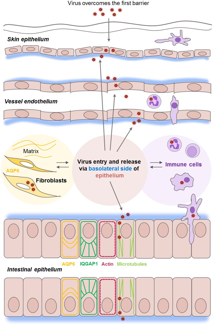
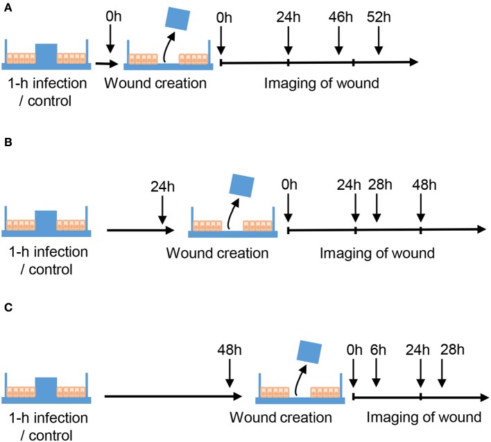
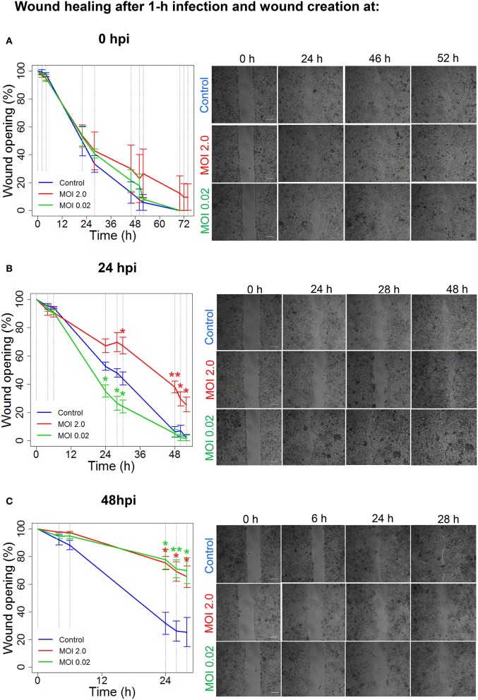
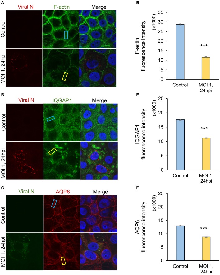
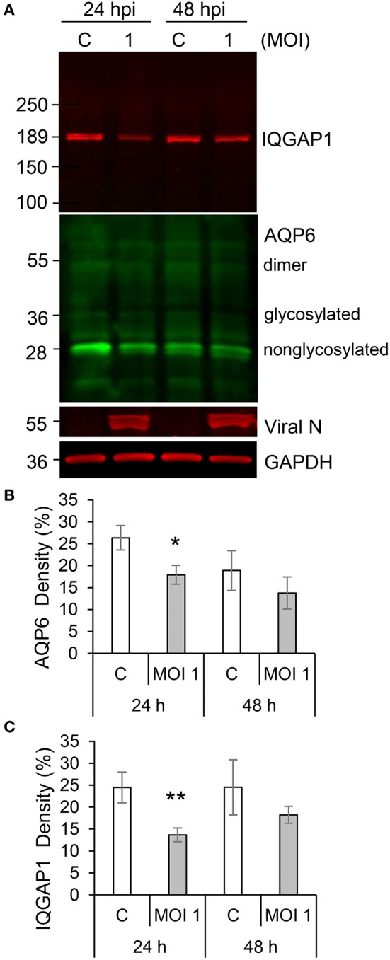
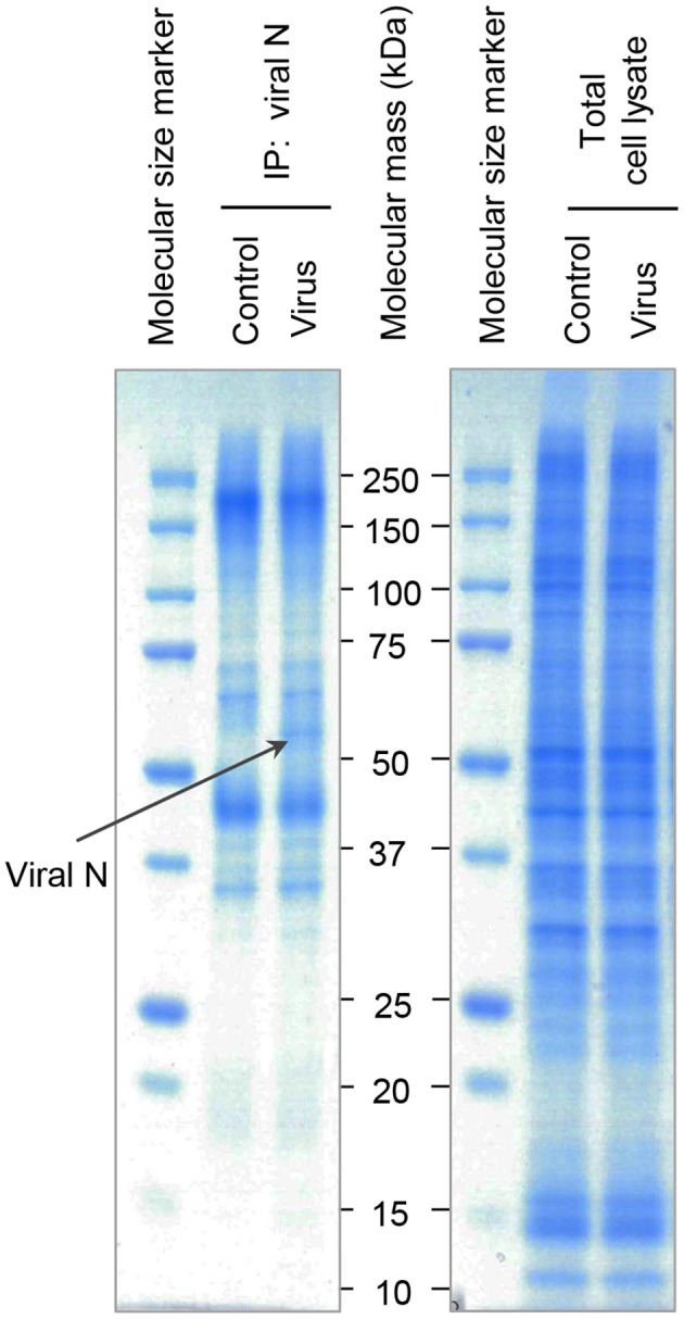
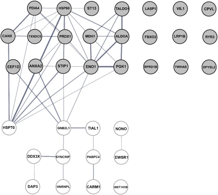
Normal epithelial and endothelial renewal and healing after bacterial and viral challenges are essential for homeostasis along the intestine and the blood and lymphatic vessels. We thus investigated whether and how virus affects migration of human epithelial cells and specifically how the nucleocapsid protein (N) modulates the cellular proteome and interactome using human Caco-2 cells in a wound-healing assay with Hazara virus as a model. Here, Hazara virus blocked cell migration in a dose- and time-dependent manner, disrupted the actin cytoskeleton and specifically reduced the expression of the IQ-motif-containing GTPase-activating protein 1 (IQGAP1) and water channel aquaporin 6 (AQP6) that regulate cytoskeletal organization, water homeostasis and vesicle communication. Moreover, in the Caco-2 cell proteome, we identified several distinct groups of molecules associating with N upon Hazara virus infection, being involved in the ensemble of important cellular processes, e.g., chaperone activity, metabolism, cellular defense against infections, cell morphology, and migration. These events do not only facilitate the virus life cycle, but they are also crucial for membrane and cytoskeleton dynamics, cellular self-renewal and wound healing, being so essential for body integrity and homeostasis.

Keywords: IQGAP1; aquaporin 6; cellular proteome; epithelial barrier; epithelial migration; homeostasis; virus infection.


Figures

Figure 1
Figure 1
Epithelial barrier functions at the front line of viral infections. Epithelial and endothelial cell monolayers (in pink) provide a series of host barriers to pathogens and other environmental agents. These possess both physical and immune (in lilac) properties. Epithelium constantly moving, renewing, and normally undergoes a wound-healing process which is controlled by an interplay between matrix, water homeostasis (in yellow), and cytoskeleton and junctions-associated proteins (ex. IQGAP1 in green). Viruses of the Nairovirus genus (red asterisk) can overcome the first host barrier to enter the body and access underlying cells. Viruses enter host cells by endocytosis, replicates, and assembles in the perinuclear regions of the cytoplasm and leaves the epithelial cell monolayers from the basolateral side (in blue), and further utilize microtubule (light green) and actin (red), the components of host cytoskeleton during the whole life cycle.
Figure 2
Figure 2
Schematic layout of wound healing assays. Caco-2 cells were cultured in μ-dishes with Ibidi inserts until monolayers were confluent. Cells were then either untreated (Control) or infected with Hazara virus at different MOI for 1 h. After 1-h infection, the cells were rinsed and maintained for 0, 24, or 48 hpi as shown in (A–C) respectively. To create a wound, the inserts were removed at 0, 24, or 48 hpi, and the cells were thereafter allowed to migrate. To monitor the wound healing, the images of cells migrating into the gap area of the wound were taken at indicated time using the benchtop microscope.
Figure 3
Figure 3
Hazara virus modulates wound healing. Wound healing assays were performed as shown in Figure 2. Cell monolayers were either untreated (Control) or infected with Hazara virus at MOI 2.0 and 0.02 for 1 h. After 1-h infection, the cells were rinsed and maintained for 0, 24, or 48 hpi, as in (A–C) respectively. To create a wound, the inserts were removed at 0, 24, or 48 hpi, and the cells were thereafter allowed to migrate as shown in images in in the right panels in (A–C), respectively. The graphs in the left panels represent quantification of wound opening, shown as percent (%) of the original gap. Values are the mean ± SE based on three independent experiments performed on separate days from different cell passages (n = 3). Significant differences were analyzed by Student's two-tailed t-test and are indicated with * or ** when P < 0.05 or P < 0.01 compared to control. Representative images of one of three independent experiments are shown in panels on the right. Bar 200 μm.
Figure 4
Figure 4
Visualization of Hazara virus, F-actin, IQGAP1 and AQP6 in epithelial cells. Caco-2 cell monolayers were infected with Hazara virus at MOI 1 for 1 h and maintained for 24 hpi. Untreated non-infected Caco-2 cell monolayers were used as a control. Samples were then fixed and stained for: (A) viral N (red) and F-actin (green); (B) viral N (red) and IQGAP1 (green); (C) viral N (green) and AQP6 (red); nuclei were labeled with DAPI (blue). Samples were analyzed by confocal microscopy. The data is from one representative of three independent experiments. Image size is 67.6 × 67.6 μm and pixel size is 0.13 μm. Quantification of fluorescence intensities for F-actin (D), IQGAP1 (E) and AQP6 (F) measured in juxta-membrane regions in cell monolayers as indicated by blue (untreated control) and yellow rectangles (virus-infected cells, MOI 1, 24 hpi) in (AC). Values in graphs represent the mean ± SE based on 3 independent experiments and 60–63 cells for each condition (same color code as in images). Significant differences were analyzed by Student's t-test and are indicated with *** when P < 0.001 compared to control.
Figure 5
Figure 5
Hazara virus affects the level of IQGAP1 and AQP6 in epithelial cells. (A) Cells were untreated controls (c) or infected with Hazara virus at MOI 1 for 1 h and maintained for 24 and 48 hpi. Immunoblots for IQGAP1, AQP6, viral N, and GAPDH. The blots are from one representative out of four independent experiments: (B,C) Quantification of blots. AQP6 and IQGAP1 levels normalized to the GAPDH control are indicated as percent (%). Values are the mean ± SE based on four independent experiments performed on separate days from different cell passages (n = 4). Significant differences were analyzed by Student's t-test and are indicated with * or ** when P < 0.05 or P < 0.01 compared to control.
Figure 6
Figure 6
SDS-PAGE analysis of N-associated immune complexes of epithelial cells. Cells were untreated (Control) or infected with Hazara virus at MOI 1 for 1 h and maintained for 24 hpi (Virus). Total cell lysates were immunoprecipitated using the antibodies against CCHFV/Hazara (viral N). N-enriched immune complexes (Left Panel) and total cell lysates as a control (Right Panel) were analyzed by SDS-PAGE and stained with Coomassie Blue. Bands were excised and N-binding cellular proteins identified by in-gel digestion and LC-MS/MS analysis as shown in Table 1. The band indicated with a black arrow represent viral N. Displayed are representative gels from one of four independent experiments.
Figure 7
Figure 7
Bioinformatic assessment of the human cellular interactome of the Hazara virus N shown in Table 1. Network nodes represent human proteins pooled into two units. The upper unit with gray nodes represent 23 proteins suggested as potential N partners upon Hazara infection. These include 10 N-associated proteins detected only in virus-infected cells and 13 proteins displayed at least 2-fold increased binding to N-enriched immune complexes upon viral infection in comparison to non-infected control (Table 1). The lower unit with white nodes represent 12 proteins displayed decreased binding to N-enriched immune complexes upon viral infection in comparison to non-infected control (Table 1). Edges represent protein-protein interactions where line thickness indicates the strength of data support. The assessment is based on STRING analysis shown in Figure S1 and LC-MS/MS analysis and quantification of alterations in cellular proteome and interactome of N shown in Table 1.
Figure 8
Figure 8
Schematic model. Epithelial wound healing process and cellular protein networks (shown as arrows) in human epithelial cells (in pink) are perturbed by Hazara virus infection. This is paralleled by disruption in the organization of actin cytoskeleton (in red) and reduction of the expression and distribution of IQGAP1 (in green) and AQP6 (in yellow), events that influence homeostasis in the intestine and along blood vessel walls and integrity of an individual.

Similar articles

Cited by

References

    1. Abbas W., Kumar A., Herbein G. (2015). The eEF1A proteins: at the crossroads of oncogenesis, apoptosis, and viral infections. Front. Oncol. 5:75. 10.3389/fonc.2015.00075 - DOI - PMC - PubMed
    1. Andersson I., Simon M., Lundkvist A., Nilsson M., Holmstrom A., Elgh F., et al. . (2004). Role of actin filaments in targeting of Crimean Congo hemorrhagic fever virus nucleocapsid protein to perinuclear regions of mammalian cells. J. Med. Virol. 72, 83–93. 10.1002/jmv.10560 - DOI - PubMed
    1. Beitz E., Liu K., Ikeda M., Guggino W. B., Agre P., Yasui M. (2006). Determinants of AQP6 trafficking to intracellular sites versus the plasma membrane in transfected mammalian cells. Biol. Cell 98, 101–109. 10.1042/BC20050025 - DOI - PubMed
    1. Benga G. (2012). On the definition, nomenclature and classification of water channel proteins (aquaporins and relatives). Mol. Aspects Med. 33, 514–517. 10.1016/j.mam.2012.04.003 - DOI - PubMed
    1. Bente D. A., Forrester N. L., Watts D. M., McAuley A. J., Whitehouse C. A., Bray M. (2013). Crimean-Congo hemorrhagic fever: history, epidemiology, pathogenesis, clinical syndrome and genetic diversity. Antiviral Res. 100, 159–189. 10.1016/j.antiviral.2013.07.006 - DOI - PubMed