

Platelet-Derived Microvesicles in Cardiovascular Diseases
- PMID: 29209618
- PMCID: PMC5702324
- DOI: 10.3389/fcvm.2017.00074
Platelet-Derived Microvesicles in Cardiovascular Diseases
Abstract
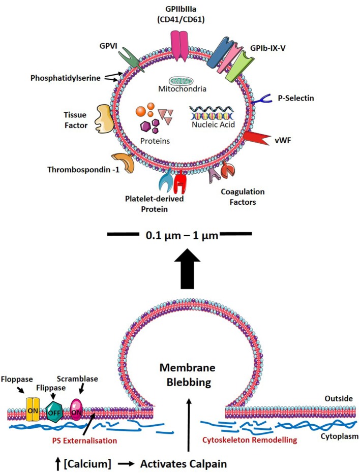
Microvesicles (MVs) circulating in the blood are small vesicles (100-1,000 nm in diameter) derived from membrane blebs of cells such as activated platelets, endothelial cells, and leukocytes. A growing body of evidence now supports the concept that platelet-derived microvesicles (PMVs), the most abundant MVs in the circulation, are important regulators of hemostasis, inflammation, and angiogenesis. Compared with healthy individuals, a large increase of circulating PMVs has been observed, particularly in patients with cardiovascular diseases. As observed in MVs from other parent cells, PMVs exert their biological effects in multiple ways, such as triggering various intercellular signaling cascades and by participating in transcellular communication by the transfer of their "cargo" of cytoplasmic components and surface receptors to other cell types. This review describes our current understanding of the potential role of PMVs in mediating hemostasis, inflammation, and angiogenesis and their consequences on the pathogenesis of cardiovascular diseases, such as atherosclerosis, myocardial infarction, and venous thrombosis. Furthermore, new developments of the therapeutic potential of PMVs for the treatment of cardiovascular diseases will be discussed.
Keywords: angiogenesis; cardiovascular disease; hemostasis; inflammation; microvesicles; platelet-derived microvesicles; therapeutic potential.
Figures

Similar articles
-
Role of Platelet-Derived Microvesicles As Crosstalk Mediators in Atherothrombosis and Future Pharmacology Targets: A Link between Inflammation, Atherosclerosis, and Thrombosis.Front Pharmacol. 2016 Aug 31;7:293. doi: 10.3389/fphar.2016.00293. eCollection 2016. Front Pharmacol. 2016. PMID: 27630570 Free PMC article. Review.
-
Platelet-derived microvesicles activate human platelets via intracellular calcium mediated reactive oxygen species release.Blood Cells Mol Dis. 2023 Jan;98:102701. doi: 10.1016/j.bcmd.2022.102701. Epub 2022 Aug 28. Blood Cells Mol Dis. 2023. PMID: 36057195
-
Association of platelet-derived microvesicles and their phenotypes with carotid atherosclerosis and recurrent vascular events in patients after ischemic stroke.Thromb Res. 2019 Apr;176:18-26. doi: 10.1016/j.thromres.2019.01.014. Epub 2019 Feb 2. Thromb Res. 2019. PMID: 30763823
-
Platelets and extracellular vesicles and their cross talk with cancer.Blood. 2021 Jun 10;137(23):3192-3200. doi: 10.1182/blood.2019004119. Blood. 2021. PMID: 33940593 Free PMC article. Review.
-
Platelet-derived microvesicles (PMVs) in cancer progression and clinical applications.Clin Transl Oncol. 2023 Apr;25(4):873-881. doi: 10.1007/s12094-022-03014-3. Epub 2022 Nov 23. Clin Transl Oncol. 2023. PMID: 36417084 Review.
Cited by
-
Hypertension Associated With Hyperlipidemia Induced Different MicroRNA Expression Profiles in Plasma, Platelets, and Platelet-Derived Microvesicles; Effects of Endothelial Progenitor Cell Therapy.Front Med (Lausanne). 2019 Dec 3;6:280. doi: 10.3389/fmed.2019.00280. eCollection 2019. Front Med (Lausanne). 2019. PMID: 31850358 Free PMC article.
-
Generation of platelet-derived microparticles through the activation of the toll-like receptor 4.Heliyon. 2019 Apr 8;5(4):e01486. doi: 10.1016/j.heliyon.2019.e01486. eCollection 2019 Apr. Heliyon. 2019. PMID: 31008410 Free PMC article.
-
Expanding applications of allogeneic platelets, platelet lysates, and platelet extracellular vesicles in cell therapy, regenerative medicine, and targeted drug delivery.J Biomed Sci. 2023 Sep 14;30(1):79. doi: 10.1186/s12929-023-00972-w. J Biomed Sci. 2023. PMID: 37704991 Free PMC article. Review.
-
Therapeutic Applications of Extracellular Vesicles for Myocardial Repair.Front Cardiovasc Med. 2021 Dec 9;8:758050. doi: 10.3389/fcvm.2021.758050. eCollection 2021. Front Cardiovasc Med. 2021. PMID: 34957249 Free PMC article. Review.
-
Platelets in cardiac ischaemia/reperfusion injury: a promising therapeutic target.Cardiovasc Res. 2019 Jun 1;115(7):1178-1188. doi: 10.1093/cvr/cvz070. Cardiovasc Res. 2019. PMID: 30906948 Free PMC article. Review.
References
Publication types
LinkOut - more resources
Full Text Sources
Other Literature Sources
Research Materials