. 2018 Apr;33(2):523-535.
doi: 10.1007/s11011-017-0162-6. Epub 2017 Dec 5.

Altered brain morphometry in 7-year old HIV-infected children on early ART

Affiliations

Altered brain morphometry in 7-year old HIV-infected children on early ART

Emmanuel C Nwosu et al. Metab Brain Dis. 2018 Apr.

Abstract

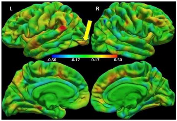
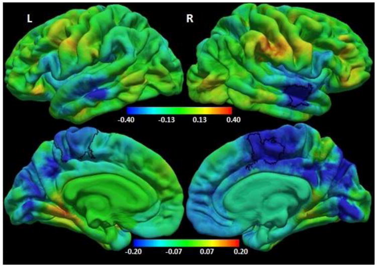
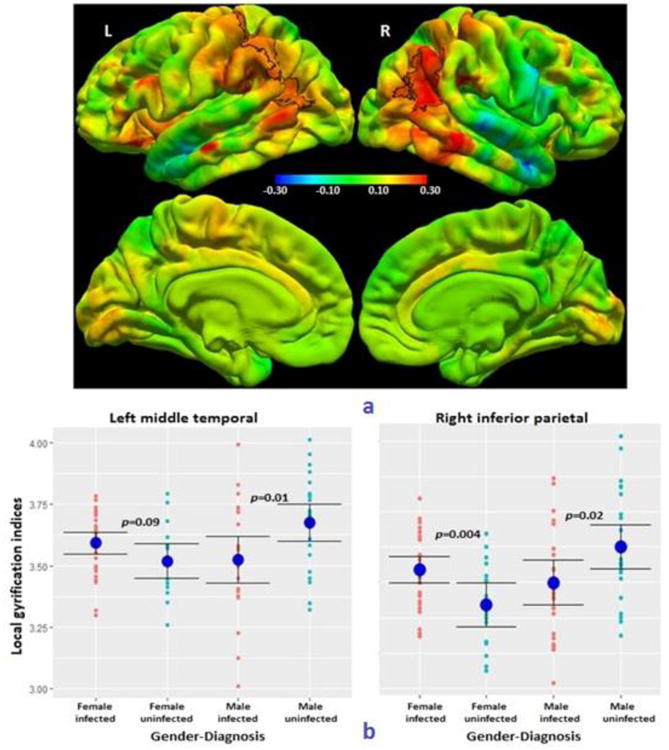
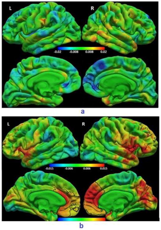
Even with the increased roll out of combination antiretroviral therapy (cART), paediatric HIV infection is associated with neurodevelopmental delays and neurocognitive deficits that may be accompanied by alterations in brain structure. Few neuroimaging studies have been done in children initiating ART before 2 years of age, and even fewer in children within the critical stage of brain development between 5 and 11 years. We hypothesized that early ART would limit HIV-related brain morphometric deficits at age 7. Study participants were 7-year old HIV-infected (HIV+) children from the Children with HIV Early Antiretroviral Therapy (CHER) trial whose viral loads were supressed at a young age, and age-matched uninfected controls. We used structural magnetic resonance imaging (MRI) and FreeSurfer ( http://www.freesurfer.net/ ) software to investigate effects of HIV and age at ART initiation on cortical thickness, gyrification and regional brain volumes. HIV+ children showed reduced gyrification compared to controls in bilateral medial parietal regions, as well as reduced volumes of the right putamen, left hippocampus, and global white and gray matter and thicker cortex in small lateral occipital region. Earlier ART initiation was associated with lower gyrification and thicker cortex in medial frontal regions. Although early ART appears to preserve cortical thickness and volumes of certain brain structures, HIV infection is nevertheless associated with reduced gyrification in the parietal cortex, and lower putamen and hippocampus volumes. Our results indicate that in early childhood gyrification is more sensitive than cortical thickness to timing of ART initiation. Future work will clarify the implications of these morphometric effects for neuropsychological function.

Keywords: Cher; Cortical thickness; Gyrification; Morphometry; Neurodevelopment; Paediatric HIV.


Conflict of interest statement

Conflict of Interest: The authors declare that they have no conflict of interest.

Figures

Fig. 1
Fig. 1
Colour map of regression coefficients for cortical thickness (mm) against group (HIV+ children/controls) controlling for sex. Positive regression coefficients (red/yellow) indicate HIV+ > controls, and negative coefficients (cyan/blue) indicate HIV+ < controls. The color bar scale applies to both lateral (top) and medial (bottom) views. The small left lateral occipital region outlined in black shows where HIV+ children have thicker cortex compared to uninfected controls at a threshold of p<0.05, with a cluster size corrected threshold of p<0.05 (Peak MNI co-ordinates 32.9, -83.4, 1.6, size 865.57 mm2).
Fig.2
Fig.2
Colour map of regression coefficients for local gyrification index (LGI) against group (HIV+ children /controls) controlling for sex. Positive regression coefficients (red/yellow) indicate HIV+ > controls, and negative coefficients (cyan/blue) indicate HIV+ < controls. Note different color bar scales for lateral (top) and medial (bottom) views. The bilateral paracentral (left: precentral/ postcentral/ paracentral/ posterior cingulate/ precuneus/ superior parietal gyri, MNI co-ordinates -6.4, -35.8, 56.5, size 1966.90mm2; right: superior frontal/ paracentral/ posterior cingulate gyri, MNI co-ordinates 11.2, 1.4, 46.1, size 1109.49 mm2) and right temporal (superior / middle temporal gyri, MNI co-ordinates 54.6, -12.3, -17.3, size 802.37mm2) regions outlined in black show where HIV+ children have smaller local gyrification indices compared to uninfected controls at a threshold of p<0.05, with a cluster size corrected threshold of p<0.05.
Fig. 3
Fig. 3
a) Colour map of regression coefficients for the interaction effect of sex on the relationship between HIV infection status and local gyrification index (LGI). The same color bar scale applies to all views. The bilateral parieto-temporal regions outlined in black show a significant sex-HIV interaction effect at a threshold of p<0.05, with a cluster size threshold of p<0.05. (Left postcentral/ superior parietal/ supramarginal/ inferior parietal/ middle temporal gyri, MNI co-ordinates -47.2, -60.0, 8.8, size 2538.16 mm2; right inferior parietal/ middle temporal/ supramarginal/ superior parietal gyri, MNI co-ordinates 45.6, -52.0, 19.7, size 2484.04 mm2) b) Plots of peak LGI (and group means ± SD) in the left and right parieto-temporal clusters, showing how the effect of HIV infection on LGIs differs in girls and boys. On the left, the peak LGI was located in the middle temporal gyrus, and on the right in the inferior parietal gyrus.
Fig. 4
Fig. 4
a) Colour map of regression coefficients for the regression of cortical thickness (mm) on age at ART initiation (weeks). The cluster outlined in black shows a right medial frontal region where older age at ART initiation was associated with thinner cortex at a threshold of p<0.05, with a cluster size threshold of p<0.05, after controlling for sex and duration of interruption. (Right rostral anterior cingulate/ medial orbitofrontal/ superior frontal/ frontal pole/ rostral middle frontal gyri, MNI co-ordinates 10.3, 51.2, 3.3, size 882.87 mm2). A similar region showed thinner cortex in After-12wk children compared to Before-12wk children. The same color bar scale applies to all views. b) Colour map of regression coefficients for LGI on age at ART initiation (weeks). Clusters outlined in black show (top) right lateral fronto-temporal (right pars orbitalis/ pars triangularis/ lateral orbitofrontal/ insula/ superior temporal, MNI co-ordinates 43.2, -0.8, -18.7, size 838.81 mm2) and (bottom) bilateral medial frontal (left: medial orbitofrontal/ rostral anterior cingulate/ caudal anterior cingulate/ superior frontal/ frontal pole/ rostral middle frontal, MNI co-ordinates -5.7, 29.6, 13.6, size 3609.06 mm2; right: caudal anterior cingulate/ rostral anterior cingulate/ medial orbitofrontal/ superior frontal/ rostral middle frontal/ frontal pole, MNI co-ordinates 14.7, 39.0, 9.2, size 5285.53 mm2) regions where older age at ART initiation was associated with greater LGIs at a threshold of p<0.05, with a cluster size threshold of p<0.05, after controlling for sex and duration of interruption. Similar regions showed greater gyrification in After-12wk children compared to Before-12wk children. Note different color bar scales for lateral (top) and medial (bottom) views.

Similar articles

Cited by

References

    1. Ackermann C, Andronikou S, Laughton B, Kidd M, Dobbels E, Innes S, van Toorn R, Cotton M. White matter signal abnormalities in children with suspected HIV-related neurologic disease on early combination antiretroviral therapy. The Pediatric infectious disease journal. 2014;33(8):e207–12. 2014. - PMC - PubMed
    1. Ackermann C, Andronikou S, Saleh MG, Laughton B, Alhamud AA, van der Kouwe A, Meintjes EM. Early Antiretroviral Therapy in HIV-Infected Children Is Associated with Diffuse White Matter Structural Abnormality and Corpus Callosum Sparing. American Journal of Neuroradiology. 2016;37(12):2363–2369. - PMC - PubMed
    1. Ances BM, Ortega M, Vaida F, Heaps J, Paul R. Independent effects of HIV, aging, and HAART on brain volumetric measures. Journal of acquired immune deficiency syndromes (1999) 2012;59(5):469. - PMC - PubMed
    1. Andronikou S, Ackermann C, Laughton B, Cotton M, Tomazos N, Spottiswoode B, Mauff K, Pettifor JM. Corpus callosum thickness on mid-sagittal MRI as a marker of brain volume: a pilot study in children with HIV-related brain disease and controls. Pediatric radiology. 2015;45(7):1016–1025. - PubMed
    1. Benjamini Y, Hochberg Y. Controlling the false discovery rate: a practical and powerful approach to multiple testing. Journal of the royal statistical society Series B (Methodological) 1995:289–300.

Publication types

Substances

LinkOut - more resources