. 2017 Dec 6;18(12):2632.
doi: 10.3390/ijms18122632.

Aberrant N-Glycosylation Profile of Serum Immunoglobulins is a Diagnostic Biomarker of Urothelial Carcinomas

Affiliations

Aberrant N-Glycosylation Profile of Serum Immunoglobulins is a Diagnostic Biomarker of Urothelial Carcinomas

Toshikazu Tanaka et al. Int J Mol Sci. .

Abstract

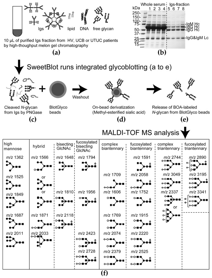
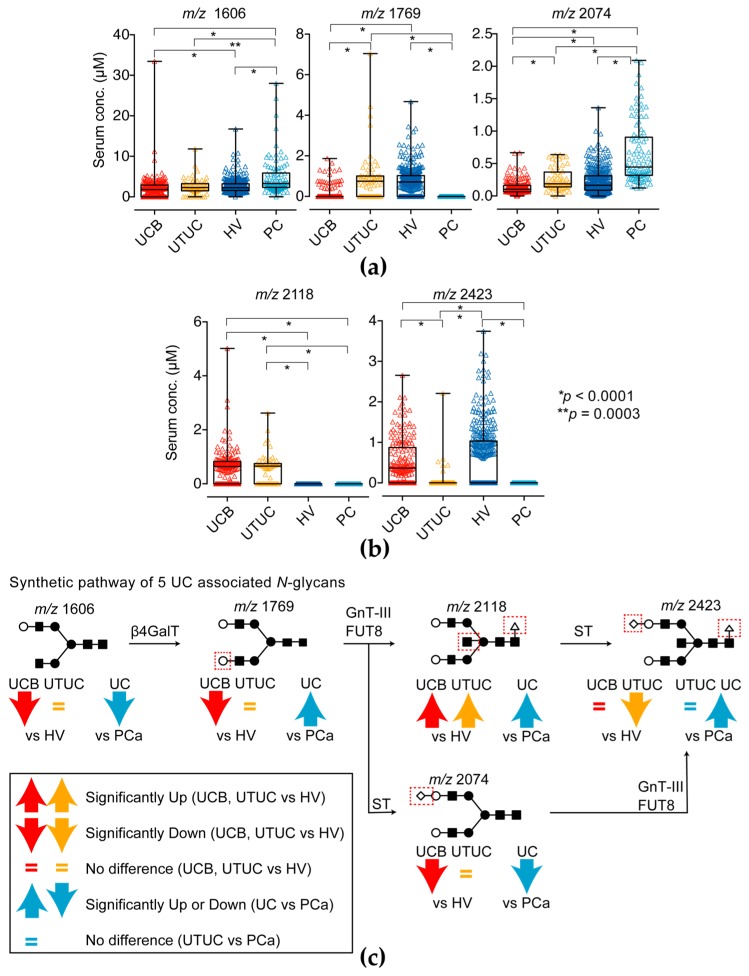
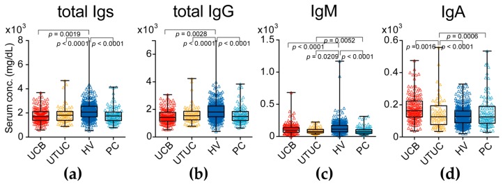
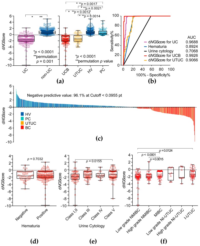
The aim of this study to determine whether the aberrant N-glycosylated serum immunoglobulins (Igs) can be applied as a diagnostic marker of urothelial carcinoma (UC). Between 2009 and 2016, we randomly obtained serum available from 237 UC and also 96 prostate cancer as other cancer controls from our serum bank and also obtained-from 339 healthy volunteers (HV)-controls obtained from community-dwelling volunteers in Iwaki Health Promotion Project. A total of 32 types of N-glycan levels on Igs were determined by high-throughput N-glycomics and analyzed by multivariable discriminant analysis. We found five UC-associated aberrant N-glycans changes on Igs and also found that asialo-bisecting GlcNAc type N-glycan on Igs were significantly accumulated in UC patients. The diagnostic N-glycan Score (dNGScore) established by combination of five N-glycans on Igs discriminated UC patients from HV and prostate cancer (PC) patients with 92.8% sensitivity and 97.2% specificity. The area under the curve (AUC) for of the dNGScore was 0.969 for UC detection that was much superior to that of urine cytology (AUC, 0.707) and hematuria (AUC, 0.892). Furthermore, dNGScore can detect hematuria and urine cytology negative patients. The dNGscore based on aberrant N-glycosylation signatures of Igs were found to be promising diagnostic biomarkers of UCs.

Keywords: N-glycomics; aberrant N-glycosylation; diagnostic biomarker; immunogloburins; upper urinary tract urothelial carcinoma; urothelial carcinoma of the bladder.


Conflict of interest statement

Japanese patent application number 2017-207525. This patent application is a domestic priority application from Japanese patent application number 2017-076018.

Figures

Figure A1
Figure A1
The general protocol for the integrated glycoblotting technique and workflow for glycoblotting-based high-throughput clinical glycan analysis. (a) Ten-microliter serum Ig fraction samples were applied to SweetBlot™ (System Instruments, Hachioji, Japan) for glycoblotting; (b) Coomassie brilliant blue (CBB)-stained band patterns on a sodium dodecyl sulfate-polyacrylamide gel for representative whole serum and purified Ig fraction samples; (c) After enzymatic N-glycan cleavage from Ig proteins, the total N-glycans released into the digestion mixture were directly mixed with BlotGlyco H beads (Sumitomo Bakelite, Co., Tokyo, Japan) to capture N-glycans; (d) After the beads were separated from other molecules by washing, sialic acid was methyl esterified; (e) These processed N-glycans were then labelled with benzyloxyamine (BOA) and released from BlotGlyco H beads; (f) Mass spectra of BOA-labelled N-glycans from Ig fractions were acquired using an Ultraflex III instrument (Bruker Daltonics, Germany). Total 32 type of N-glycans were identified in Igs fraction. Identified N-glycan structures are indicated by monosaccharide symbols: white circles, galactose (Gal); black circles, mannose (Man); black squares, N-acetylglucosamine (GlcNAc); black triangle, fucose (Fuc) and black diamonds, sialic acid.
Figure 1
Figure 1
Five UC-associated N-glycan levels of immunoglobulins (Igs) fractions. (a) Complex biantennary-type N-glycan (m/z 1606, 1769 and 2074) levels and (b) fucosylated bisecting GlcNAc-type N-glycan (m/z 2118 and 2423) of the Ig fractions in the UC, HV and PC groups. The results shown are representative of three independent experiments. Intergroup differences were statistically compared using the Mann–Whitney U-test for non-normally distributed models; (c) Synthetic pathway of candidate N-glycans in the present study. Red up- and downward arrows indicates significantly changed in UC compare with HV group. Cyan up- and downward arrows indicates significantly changed in UC compare with PC group. The equals mark indicates that it did not significantly change. N-glycan structures are indicated by monosaccharide symbols: white circles, galactose (Gal); black circles, mannose (Man); black squares, N-acetylglucosamine (GlcNAc); black triangle, fucose (Fuc) and black diamonds, sialic acid. β4GalT: β1,4-galactosyltransferase, GnT-III: N-acetylglucosaminyltransferase-III, FUT8: fucosyltransferase 8, ST: sialyltransferase.
Figure 2
Figure 2
Serum immunoglobulin (Ig) levels in UC (UC of the bladder (UCB) and upper urinary tract UC (UTUC)) patients and healthy volunteers (HVs) and prostate cancer (PC). (a) Serum total Igs (sum of IgG1-4, IgM and IgA) levels in the UC, HV and PC groups; (b) Serum total IgG (IgG1-4) levels in the UC, HV and PC groups; (c) Serum IgM levels in the UC, HV and PC groups; (d) Serum IgA levels in the UC, HV and PC groups. The results shown are representative of three independent experiments. Intergroup differences were statistically compared using the Mann–Whitney U-test for non-normally distributed models.
Figure 3
Figure 3
Clinical significance of diagnostic N-glycan score. (a) The diagnostic N-glycan score (dNGScore) level was significantly higher in the UC (UCB and UTUC) group than in the HV and PC group; (b) ROC curve analysis of dNGScore, hematuria and urine cytological results for detection of UC (UCB and UTUC); (c) Waterfall plot of dNGScore; (d) Association between dNGScore level and hematuria status; (e) Association between dNGScore level and urine cytology; (f) Association between dNGScore level and tumor invasiveness. NMIBC: non-muscle invasive bladder cancer, MIBC: muscle invasive bladder cancer, NI-UTUC: noninvasive UTUC, I-UTUC: invasive UTUC. Intergroup differences were statistically compared using the Mann–Whitney U-test for non-normally distributed models and performed permutation test.

References

    1. Park J.C., Hahn N.M. Bladder cancer: A disease ripe for major advances. Clin. Adv. Hematol. Oncol. 2014;12:838–845. - PubMed
    1. Munoz J.J., Ellison L.M. Upper tract urothelial neoplasms: Incidence and survival during the last 2 decades. J. Urol. 2000;164:1523–1525. doi: 10.1016/S0022-5347(05)67019-X. - DOI - PubMed
    1. Siegel R., Naishadham D., Jemal A. Cancer statistics, 2012. CA Cancer J. Clin. 2012;62:10–29. doi: 10.3322/caac.20138. - DOI - PubMed
    1. Inman B.A., Tran V.T., Fradet Y., Lacombe L. Carcinoma of the upper urinary tract: Predictors of survival and competing causes of mortality. Cancer. 2009;115:2853–2862. doi: 10.1002/cncr.24339. - DOI - PubMed
    1. Cowan N.C. CT urography for hematuria. Nat. Rev. Urol. 2012;9:218–226. doi: 10.1038/nrurol.2012.32. - DOI - PubMed

LinkOut - more resources