. 2017 Dec 6;6(4):102.
doi: 10.3390/antiox6040102.

In Vitro and In Vivo Antioxidant and Anti-Hyperglycemic Activities of Moroccan Oat Cultivars

Affiliations

In Vitro and In Vivo Antioxidant and Anti-Hyperglycemic Activities of Moroccan Oat Cultivars

Ilias Marmouzi et al. Antioxidants (Basel). .

Abstract

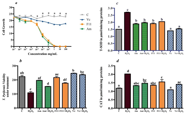
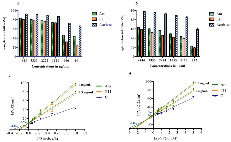
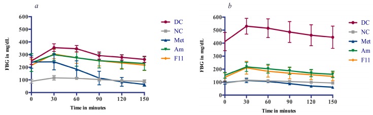
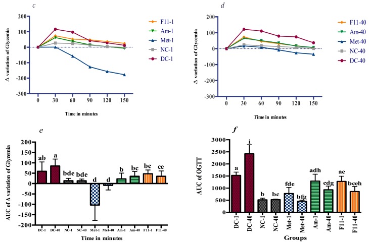
Improvement of oat lines via introgression is an important process for food biochemical functionality. This work aims to evaluate the protective effect of phenolic compounds from hybrid Oat line (F11-5) and its parent (Amlal) on hyperglycemia-induced oxidative stress and to establish the possible mechanisms of antidiabetic activity by digestive enzyme inhibition. Eight phenolic acids were quantified in our samples including ferulic, p-hydroxybenzoic, caffeic, salicylic, syringic, sinapic, p-coumaric and chlorogenic acids. The Oat extract (2000 mg/kg) ameliorated the glucose tolerance, decreased Fasting Blood Glucose (FBG) and oxidative stress markers, including Superoxide dismutase (SOD), Catalase (CAT), Glutathione peroxidase (GPx), Glutathione (GSH) and Malondialdehyde (MDA) in rat liver and kidney. Furthermore, Metformin and Oat intake prevented anxiety, hypercholesterolemia and atherosclerosis in diabetic rats. In vivo anti-hyperglycemic effect of Oat extracts has been confirmed by their inhibitory activities on α-amylase (723.91 μg/mL and 1027.14 μg/mL) and α-glucosidase (1548.12 μg/mL & 1803.52 μg/mL) enzymes by mean of a mixed inhibition.

Keywords: anti-hyperglycemic; digestive enzyme; hybrid Oat; streptozotocin-nicotinamide.


Conflict of interest statement

The authors declare no conflict of interest.

Figures

Figure 1
Figure 1
Oat protective effect on antioxidant enzymes under oxidative stress in T. pyriformis. (a) Oat and Vitamin C effect on T. pyriformis growth. (b) T. pyriformis viability under H2O2 treatment in the presence of extracts. (c) T-SOD levels T. pyriformis cultures. (d) CAT levels T. pyriformis cultures, Data are reported to mean (n = 3) ± SD. Values not sharing a common letter (a–d) differs significantly at p < 0.05. (*) differ significantly from the control.
Figure 2
Figure 2
α-amylase and α-glucosidase inhibitory activities. (a) α-amylase inhibitory activities of Oat extracts. (b) α-glucosidase inhibitory activities of Oat extracts. (c) Lineweaver burk plot of Oat α-amylase inhibition. (d) Lineweaver burk plot of Oat α-glucosidase inhibition, Data are reported to mean (n = 3) ± SD.
Figure 3
Figure 3
Oral glucose tolerance before and after treatment period. (a) OGTT of the first day 1; (b) OGTT of the day 40; (c) Δ variation of glycaemia during OGTT-Day 1; (d) Δ variation of glycaemia during OGTT-Day 40; (e) AUC of Δ variation of glycaemia during OGTT challenge; (f) AUC of OGTT, Data are reported to mean (n = 8) ± SD. Values not sharing a common letter (a–i) differs significantly at p < 0.05.
Figure 3
Figure 3
Oral glucose tolerance before and after treatment period. (a) OGTT of the first day 1; (b) OGTT of the day 40; (c) Δ variation of glycaemia during OGTT-Day 1; (d) Δ variation of glycaemia during OGTT-Day 40; (e) AUC of Δ variation of glycaemia during OGTT challenge; (f) AUC of OGTT, Data are reported to mean (n = 8) ± SD. Values not sharing a common letter (a–i) differs significantly at p < 0.05.
Figure 4
Figure 4
Effect of diabetes and oat treatment on histological alterations in the pancreas. Representative photomicrographs of histological alterations stained with hematoxylin and eosin at ×400 magnification. Normal control (a); Diabetic control (b); Metformin (c); Amlal (d); F11-5 (e).

References

    1. Rains J.L., Jain S.K. Oxidative stress, insulin signaling, and diabetes. Free Radic. Biol. Med. 2011;50:567–575. doi: 10.1016/j.freeradbiomed.2010.12.006. - DOI - PMC - PubMed
    1. Robertson R.P. Chronic oxidative stress as a central mechanism for glucose toxicity in pancreatic islet beta cells in diabetes. J. Biol. Chem. 2004;279:42351–42354. doi: 10.1074/jbc.R400019200. - DOI - PubMed
    1. Wold L.E., Asli F.C., Jun R. Oxidative stress and stress signaling: Menace of diabetic cardiomyopathy. Acta Pharmacol. Sin. 2005;26:908–917. doi: 10.1111/j.1745-7254.2005.00146.x. - DOI - PubMed
    1. Evans J.L., Goldfine I.D., Maddux B.A., Grodsky G.M. Are Oxidative Stress—Activated Signaling Pathways Mediators of Insulin Resistance and β-Cell Dysfunction? Diabetes. 2003;52:1–8. doi: 10.2337/diabetes.52.1.1. - DOI - PubMed
    1. Henriksen J.E., Diamond-Stanic M.K., Marchionne E.M. Oxidative stress and the etiology of insulin resistance and type 2 diabetes. Free Radic. Biol. Med. 2011;51:993–999. doi: 10.1016/j.freeradbiomed.2010.12.005. - DOI - PMC - PubMed

LinkOut - more resources