. 2017 Dec 14;552(7684):194-199.
doi: 10.1038/nature25016. Epub 2017 Dec 6.

Alcohol-abuse drug disulfiram targets cancer via p97 segregase adaptor NPL4

Affiliations

Alcohol-abuse drug disulfiram targets cancer via p97 segregase adaptor NPL4

Zdenek Skrott et al. Nature. .

Abstract

Cancer incidence is rising and this global challenge is further exacerbated by tumour resistance to available medicines. A promising approach to meet the need for improved cancer treatment is drug repurposing. Here we highlight the potential for repurposing disulfiram (also known by the trade name Antabuse), an old alcohol-aversion drug that has been shown to be effective against diverse cancer types in preclinical studies. Our nationwide epidemiological study reveals that patients who continuously used disulfiram have a lower risk of death from cancer compared to those who stopped using the drug at their diagnosis. Moreover, we identify the ditiocarb-copper complex as the metabolite of disulfiram that is responsible for its anti-cancer effects, and provide methods to detect preferential accumulation of the complex in tumours and candidate biomarkers to analyse its effect on cells and tissues. Finally, our functional and biophysical analyses reveal the molecular target of disulfiram's tumour-suppressing effects as NPL4, an adaptor of p97 (also known as VCP) segregase, which is essential for the turnover of proteins involved in multiple regulatory and stress-response pathways in cells.


Conflict of interest statement

R.J.D. is a founder and consultant for Cleave Biosciences. The remaining authors declare no competing financial interests. Readers are welcome to comment on the online version of the paper.

Figures
Extended Data 1
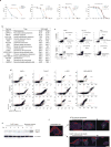
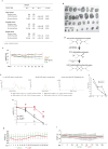
Extended Data 1. Anti-cancer effects of DSF: epidemiological and pre-clinical data
a) Summary of Hazard Ratios (HR) and 95% confidence intervals (CI) for cancer-specific mortality among Danish cancer patients comparing continuing and previous users of disulfiram (DSF) for selected types of cancer (for statistical analysis and definitions of DSF exposure categories, see Methods); b) Photographs of subcutaneously growing human MDA-MB-231 tumours extracted from mice at day 32; c) Time-course diagram of mice weight (n=8 animals per group, mean, SD); d) Model of CuET formation during metabolic processing of orally administered DSF in the human body. e) Examples of Mass-spectrometry Spectra of CuET expressed as peaks of 4 MRM transitions in murine serum after CuET spikes, compared to orally applied DSF (50 mg/kg) (representative of two independent experiments). f) Pharmacokinetic analysis of CuET levels in murine serum after orally applied DSF (50 mg/kg) (mean, SD, n=2 animals for each time point). g) Effect of DTC and CuET on MDA-MB-231 cells analysed by colony formation assay (CFA) (3 independent experiments, means linked). h) Time-course diagram of weight in CuET- and vehicle-treated mice (n=10 animals per group, mean, SD); i) Extended time-course diagram of weight in CuET- and vehicle-treated mice (n=10 animals per group, mean, SD).
Extended Data Figure 2
Extended Data Figure 2. CuET as the major anticancer metabolite of DSF
a) CuET cytotoxicity measured by a Colony formation assay (CFA) in human cell lines derived from breast (mean and SD from 3 independent experiments), lung, colon and prostate carcinomas (presented 2 independent biological experiments for each cell line); b) IC50 values from two independent biological experiments documenting differential CuET-induced cytotoxicity across a panel of cancer and non-cancerous cell lines (48h treatment); c) Analysis of AnexinV signal in AMO-1 cells exposed to toxic doses of NMS873 (5μM, 16h) or CuET (100nM, 16h) and in U-2OS cell exposed to toxic doses of NMS873 (10μM, 16h) or CuET (1μM, 16h); d) Analysis of Caspase 3/7 activity in selected cell lines after apoptosis induction by NMS873 (AMO-1: 6h, 5μM, Capan1: 16h, 10μM, U-2OS: 16h, 10μM, MDA-MB-231: 24h,10μM) or CuET (AMO-1: 16h, 100nM, Capan1: 16h, 250nM, U-2OS: 16h, 1μM, MDA-MB-231: 24h, 1 μM) e) Absence of cleaved Parp1 after toxic dose of CuET in U-2OS cells, compared to etoposide treatment as a positive control; f) Analysis of Cytochrome C (in red) release from mitochondria in U-2OS cells during cell death induced either by the positive control staurosporin (STS, 1μM) compared to cell death induced by CuET (1μM) (blue=DAPI signal). Panels c-f are representative of two independent biological experiments.
Extended Data Figure 3
Extended Data Figure 3. CuET-induced proteasome inhibition-like response is not due to proteasome inhibition
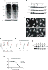
a) Kinetics of poly-Ub-protein accumulation in U-2OS cells treated with CuET or the proteasome inhibitor BTZ; b) CuET treatment (1,5h) induces rapid deubiquitylation of ubiquitylated histone H2A (uH2A) similarly to proteasome inhibitors BTZ or MG132 (U-2OS cells); c) CuET treatment (1,5h) induces rapid cytoplasmic accumulation of poly-ubiquitylated proteins (FK2 antibody staining, U-2OS cells); analogous to BTZ and MG132; d) 20S proteasome activity is not inhibited by CuET as examined in live MDA-MB-231 cells or e) in lysates from MDA-MB-231 cells (mean and SD from 4 independent experiments); f) CuET treatment (1μM, 6h) does not cause accumulation of p53 in the presence of Dicoumarol (300 μM) in MCF7 cells; g) In-vitro 26S proteasome function measured as Rpn11 deubiquitylation activity, is not inhibited by CuET; 1,10 phenantroline (1,10-OPT) served as a positive control (representative of three independent experiments). Panels a-c, f are representative of two independent experiments.
Extended Data Figure 4
Extended Data Figure 4. CuET inhibits the p97 pathway and induces cellular unfolded protein response (UPR)
a) Proteasome inhibitor MG132-treated cells (5μM, 6h) accumulate both forms of NRF1 (120- and 110-KDa bands, upper and lower arrows, respectively) while CuET-treated cells (1μM, 6h) accumulate only the non-cleaved 120-KDa form; b) Inhibition of the NRF1 cleavage process (appearance of the lower band) by CuET and NMS873 (a p97 inhibitor; 5μM) in mouse NIH3T3 cells co-treated with the proteasome inhibitor MG132 (5μM for 6h); c) Time-course example images from a FRAP experiment the quantitative analysis of which is shown in Fig. 2g (U-2OS cells, blue boxes mark areas before bleaching, arrows after bleaching); d) U-2OS cells pre-extracted by TritonX and stained for K-48-polyUb. The Ab signal intensities for cells treated with DMSO, BTZ (1μM), NMS873 (10μM) and CuET (1μM) are analysed by microscopy-based cytometry and plotted below; e) Western blot analysis of accumulated poly-Ub proteins in ultracentrifugation-separated microsomal fraction from U-2OS cells treated by mock, CuET (1μM), NMS873 (10μM) or BTZ (1μM) for 3h; f) UPR in U-2OS and MDA-MB-231 cell lines induced by 6-h treatment with CuET (various concentrations) or positive controls (NMS873 5μM, tunicamycin 2μg/ml, thapsigargin 1μM) manifested by increased levels of Xbp1s, ATF4 and p-eIF2a. Panels a-f are representative of two independent experiments.
Extended Data Figure 5
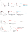
Extended Data Figure 5. CuET kills bortezomib-resistant cells
a) Bortezomib/Carfilzomib (BTZres/CFZres)-adapted and non-adapted AMO-1 human myeloma cells are similarly sensitive to treatment with CuET; b) Bortezomib/Carfilzomib (BTZres/CFZres)-adapted and non-adapted ARH77 human plasmocytoma cells are similarly sensitive to treatment with CuET; c) Bortezomib (BTZres)-adapted and non-adapted RPMI8226 human myeloma cells are similarly sensitive to treatment with CuET; d) Human myeloma cells derived from a BTZ-resistant patient show CuET sensitivity comparable with myeloma cells derived from a BTZ-sensitive patient. The charts in (ac) show three independent experiments (means linked), panel (d) shows two independent experiments.
Extended Data Figure 6
Extended Data Figure 6. CuET targets NPL4, causing NPL4’s immobilization and nuclear clustering
a) CuET (1μM) does not inhibit ATPase activity of p97; NMS873 (5μM) was used as a positive control (mean, SD from 4 independent experiments); b) Western blotting analysis documenting levels of ectopic p97-GFP, NPL4-GFP and UFD1-GFP in stable U-2OS-derived cell lines used for the CuET-treatment rescue and cluster formation experiments; c) Ectopic expression of NPL4-GFP alleviates CuET-induced (125nM, 4h) accumulation of poly-Ub proteins in U-2OS cells; d) Distribution of NPL4 nuclear clusters relative to chromatin in cells treated by CuET (1μM, 2h) (Scale bar = 2 micrometres); e) Schematic representation of site-directed mutagenesis within the amino acid sequence of the putative zinc finger domain of NPL4; f) Isothermal calorimetry curve documenting the lack of CuET binding to purified MUT-NPL4 protein; g) DARTS analysis of recombinant NPL4 proteins: differential pronase-mediated proteolysis after CuET addition is apparent for WT-NPL4 but not for MUT-NPL4; detected by either Silver-stained SDS-PAGE (the most prominent differential bands are marked by red dots) or by blotting with an anti-NPL4 polyclonal antibody; h) Viability of cells expressing a doxycycline-inducible MUT-NPL4-GFP, treated with CuET for 48 h (3 independent experiments, means are linked); i) Accumulation of K48-ubiquitinated proteins and activation of UPR in cells expressing the doxycycline-inducible MUT-NPL4-GFP. Panels b-d, f, g and i are representative of two independent experiments.
Extended Data Figure 7
Extended Data Figure 7. Immobilized NPL4 forms insoluble protein aggregates
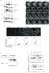
a) NPL4-GFP aggregates induced by CuET treatment (1μM for 3h) do not co-localize with nuclear speckles (stained by SC-35 antibody) or nucleoli (visible as DAPI-negative nuclear signal); b) NPL4-GFP nuclear aggregates induced by CuET (1μM, 3h) are excluded from chromatin in early prometaphase U-2OS cells; c) Co-localization of spontaneous MUT-NPL4-GFP aggregates with Sumo 2/3, K-48 polyubiquitin and TDP43 (U-2OS cells, pre-extracted); d) NPL4-GFP aggregates are formed independently of ubiquitylations, as documented on CuET (1μM, 3h) treated cells pre-treated with a chemical UBA1 inhibitor (MLN7243, 10 μM for 1h); The lack of the cellular FK2 staining for ubiquitylated proteins validates the efficacy of the MLN7243 inhibitor; e) Co-localization of FK2 signal with the spontaneous MUT-NPL4-GFP aggregates (U-2OS cells, pre-extracted); f) Analysis of p97 in CuET-induced (1μM for 3h) NPL4-GFP aggregates (U-2OS cells, pre-extracted); g) Analysis of p97 in spontaneous MUT-NPL4-GFP aggregates (U-2OS cells, pre-extracted). Panels a-g are representative of two independent biological experiments.
Extended Data Figure 8
Extended Data Figure 8. NPL4 aggregation immobilizes the p97 binding partner and induces global cellular heat shock response (HSR)
a) Immobilization of selected proteins in Triton X100-resistant pellet fractions of CuET-treated (1μM, 3h) U-2OS cells; b) Immobilization of selected proteins in Triton X100-resistant pellet fractions from U-2OS cells expressing doxycycline-inducible MUT-NPL4-GFP (48h after induction); c) CuET dose-dependent immobilization of p97 in Triton X100-pre-extracted MDA-MB-231 cells (3h); d) Immunohistochemical staining documenting non-extractable p97 in MDA-MB-231 xenografts from mice treated by DSF or DSF+gluCu, compared to vehicle; e) HSR after CuET (8h treatment) manifested by various HSR markers detected by Western blotting of U-2OS cell extracts; f) HSR markers in U-2OS cells expressing doxycycline-inducible MUT-NPL4-GFP (24h after induction). Panels a-f are representative of two independent biological experiments.
Figure 1
Figure 1. Tumour-suppressing effects of DSF and CuET
a) Effects of per-oral DSF and gluCu on subcutaneous growth of MDA-MB-231 tumours (n=8 mice/group, mean, SD); b) CuET levels in mouse tumours and tissues (n=5 tissues, n=10 tumours, mean); c) CuET levels in human plasma after DSF treatment (n=9 patients); d) Toxicity of DTC and CuET in MDA-MB-231 cells (24h, 3 experiments, means linked); e) Effect of CuET on subcutaneous growth of MDA-MB-231 tumours in mice (n=20 tumours, mean, SD); f) Survival of CuET- vs vehicle-treated mice with implanted AMO-1 xenografts (n=10 animals/group, mean, SD, log-rank test).
Figure 2
Figure 2. CuET inhibits p97 segregase-dependent protein degradation
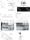
a) CuET causes accumulation of poly-ubiquitylated proteins (MCF7 cells); b) TNFα-induced IκB degradation is compromised after 1-h treatment with CuET or BTZ; c) Dose-dependent inhibition of Ub-(G76V)-GFP degradation by CuET (3 h, HeLa cells, 3 experiments, means linked); d) HIF1α levels after 2-h treatments with MG132 (5μM), CuET (1μM), BTZ (1μM) (HeLa cells). e) Differential impact of BTZ (1μM), CuET (1μM) and DBeQ (10μM) on Cdc25A vs HIF1α in MG132-pretreated (4h, 5μM), cycloheximide (1h, 50μg/ml)-exposed HeLa cells (see Methods); f) BTZ (8h, 1μM) induces NRF1 120KDa (upper arrow) and 110KDa (lower arrow) forms; while CuET (8h, 0.5μM) only the non-cleaved 120KDa form (NIH3T3 cells); g) FRAP quantification in U-2OS-Ub-GFP cells: slower mobility of accumulated cytoplasmic GFP-Ub after 2h pre-treatment with NMS873 (10μM), CuET (1μM) or BTZ (1μM) (relative mean signal of the bleached region from 12 cells per treatment). Panels a, b, d-g are representative of two independent biological experiments.
Figure 3
Figure 3. CuET binds and immobilizes NPL4
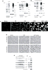
a) Ectopic NPL4-GFP, but not p97-GFP or UFD1-GFP rescues CuET toxicity (24h, U-2OS cells, mean, SD, 3 experiments); b) CuET (1μM) induces intranuclear clustering of NPL4-GFP, but not p97-GFP or UFD1-GFP; c) CuET(1μM, 2h)-induced immobilization of NPL4-GFP (FRAP, U-2OS cells, blue boxes: areas before bleaching, arrows: after bleaching); d) NPL4 enrichment in TritonX100-insoluble fractions after CuET (1μM) treatment. e) Immunohistochemistry highlights non-extractable NPL4: MDA-MB-231 tumours from mice treated by DSF or DSF+gluCu; f) Isothermal calorimetry: CuET binds to purified WT-NPL4; g) Spontaneous intranuclear clustering and immobilization of MUT-NPL4-GFP (FRAP, U-2OS cells, blue boxes: areas before bleaching, arrows: after bleaching). Scale bars=10μm. Panels b-g are representative of two independent experiments.
Figure 4
Figure 4. NPL4 protein aggregation triggers HSR
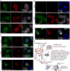
a) NPL4-GFP co-localizes with Sumo2/3, K-48 poly-Ub and TDP43 (U-2OS cells, CuET 1μM, 3h, pre-extracted); b) NPL4-GFP co-localizes with HSP70 in mock- and CuET-treated U-2OS cells (1μM, 3h, pre-extracted); c) MUT-NPL4-GFP co-localizes with HSP70 (U-2OS cells, pre-extracted); d) CuET-induced HSF1 stress bodies (1 μM, 3h, U-2OS-NPL4-GFP cells); e) HSF1 stress bodies in U-2OS cells expressing MUT-NPL4-GFP; f) Model of DSF anticancer activity in patients. All scale bars=10μm. Panels a-e are representative of two independent experiments.

Comment in

References

    1. Collins FS. Mining for therapeutic gold. Nat Rev Drug Discov. 2011;10(6):397. - PMC - PubMed
    1. Pantziarka P, et al. The Repurposing Drugs in Oncology (ReDO) Project. Ecancermedicalscience. 2014;8:442. - PMC - PubMed
    1. Iljin K, et al. High-throughput cell-based screening of 4910 known drugs and drug-like small molecules identifies disulfiram as an inhibitor of prostate cancer cell growth. Clin Cancer Res. 2009;15(19):6070–6078. - PubMed
    1. Cvek B. Nonprofit drugs as the salvation of the world’s healthcare systems: the case of Antabuse (disulfiram) Drug Discov Today. 2012;17(9–10):409–412. - PubMed
    1. Shen ML, et al. Determination of in vivo adducts of disulfiram with mitochondrial aldehyde dehydrogenase. Biochem Pharmacol. 2001;61(5):537–545. - PubMed

Publication types

MeSH terms