. 2017 Dec 5;21(10):2696-2705.
doi: 10.1016/j.celrep.2017.11.046.

Structure and Function of an Actin-Based Filter in the Proximal Axon

Affiliations

Structure and Function of an Actin-Based Filter in the Proximal Axon

Varuzhan Balasanyan et al. Cell Rep. .

Abstract

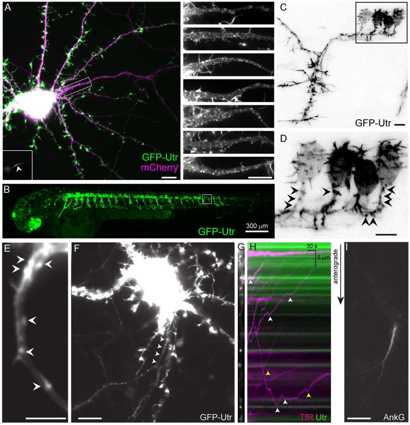
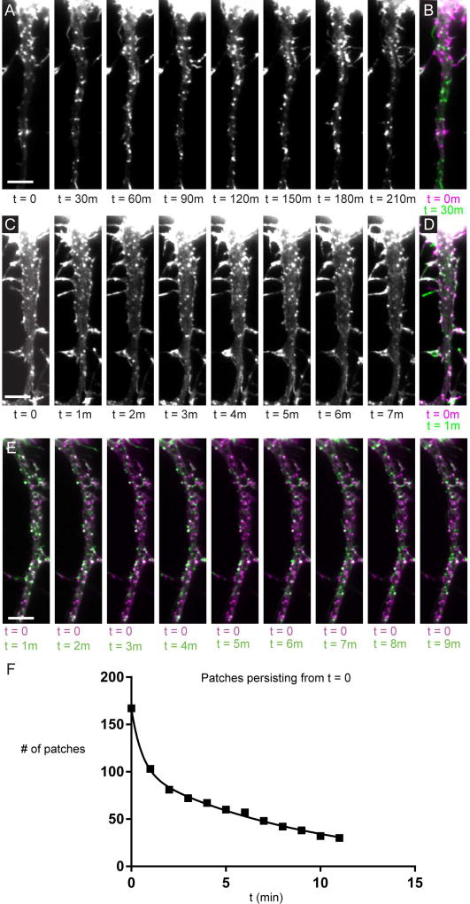
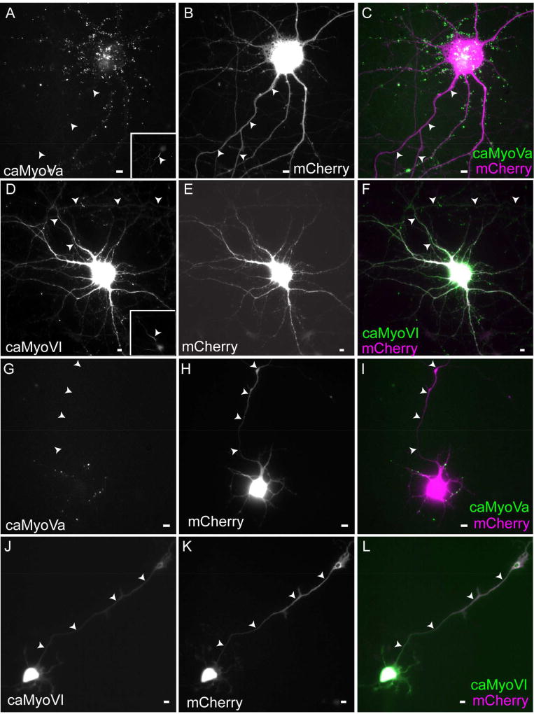
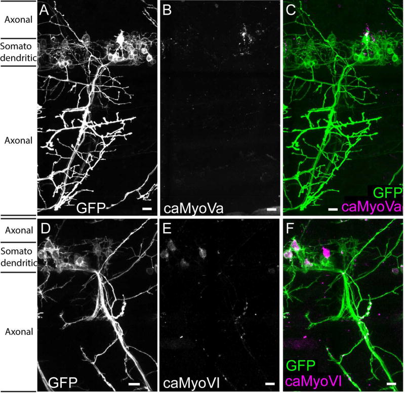
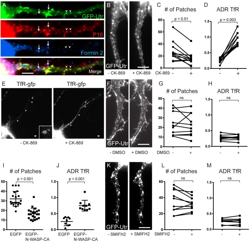
The essential organization of microtubules within neurons has been described; however, less is known about how neuronal actin is arranged and the functional implications of its arrangement. Here, we describe, in live cells, an actin-based structure in the proximal axon that selectively prevents some proteins from entering the axon while allowing the passage of others. Concentrated patches of actin in proximal axons are present shortly after axonal specification in rat and zebrafish neurons imaged live, and they mark positions where anterogradely traveling vesicles carrying dendritic proteins halt and reverse. Patches colocalize with the ARP2/3 complex, and when ARP2/3-mediated nucleation is blocked, a dendritic protein mislocalizes to the axon. Patches are highly dynamic, with few persisting longer than 30 min. In neurons in culture and in vivo, actin appears to form a contiguous, semipermeable barrier, despite its apparently sparse distribution, preventing axonal localization of constitutively active myosin Va but not myosin VI.


Figures

Figure 1
Figure 1
Actin patches are found in the proximal axons of live neurons and mark positions where vesicles carrying dendritic proteins halt and reverse. (A) 15 DIV cortical neuron in dissociated culture expressing the actin label GFP-Utr (green) and mCherry (purple). The proximal axon is contained within the boxed area. (Inset, lower left) Ankyrin G labeling of the proximal axon (arrowhead). (Right) Upper panel shows magnified image of boxed area. Lower panels show similar areas from other cortical neurons in culture expressing GFP-Utr under similar conditions. N = 20 neurons, 3 cultures. (B) Live 2 dpf zebrafish expressing GFP-Utr in motoneurons. N = 295 cells, 4 fish. (C, D) Higher magnification of area shown in boxed region of (B, C). (D) Arrowheads point to patches of actin in the proximal axon. (E) Proximal axon of 3 DIV cortical neuron expressing GFP-Utr showing actin patches (arrowheads). N = 15 neurons, 4 cultures. (F) 14 DIV cortical neuron in culture expressing TfR-mCherry (arrowheads point to the proximal axon). (G) Straightened image of proximal axon showing GFP-Utr labeling of actin patches. (H) Kymograph of vesicles carrying TfR-mCherry (purple) and actin patches (green). White arrowheads indicate places where TfR containing vesicles halted near actin patches, yellow arrowheads indicate places where halting took place away from patches. (I) Ankyrin G labeling of the axon initial segment of neuron in (F–H). Scale bar 10 µm unless otherwise indicated. See also Figures S1, S2, Movies S1, S2.
Figure 2
Figure 2
Actin patches are dynamic. (A) A series of timelapse images of the proximal axon of a cortical neuron expressing GFP-Utr taken 30 minutes apart. (B) Merge of the image at t = 0 (purple) and t = 30 minutes (green). White puncta represent patches that have persisted, purple puncta represent patches that have disappeared or moved from that location. Green puncta represent patches that have appeared or moved to that location. (C) Similar images to those in (A) taken 1 minute apart. (D) Merge of image from (C) at t = 0 (purple) and t = 1 minute (green). (E) Merge of timelapse images of GFP-Utr at t = 0 (purple) and t = 1 to 11 minutes (green). (F) Graph of the number of patches that have persisted from t = 0 over time. Solid line corresponds to the fit of 2 exponentials, Tau 1 = 0.7 +/− 0.1 minutes, Tau 2 = 6.5 +/− 0.4 minutes, R2 = 0.9979. Roughly 40% of patches have kinetics corresponding to the shorter Tau, the rest have kinetics corresponding to the longer Tau. Results are a compilation of data from 3 neurons, 3 separate cultures. Scale bar 5 µm. See also Figure S3, Movies S3–S6.
Figure 3
Figure 3
Constitutively active Myosin Va, but not Myosin VI, is excluded from the axon in cultured neurons. (A–C) caMyoVa expressed in a 16 DIV cortical neuron for 24 hours is localized exclusively in the somatodendritic compartment. Inset shows Ankyrin G staining (n = 9 neurons, 2 cultures). (D–F) caMyoVI expressed in a 16 DIV cortical neuron for 24 hours is localized to both the axonal and somatodendritic compartments (n = 12 neurons, 4 cultures). (G–I) Somatodendritic localization of caMyoVa expressed in a 3 DIV cortical neuron (n = 10 neurons, 3 cultures). (J–L) Somatodendritic and axonal localization of caMyoVI expressed in a 3 DIV cortical neuron (n = 9 neurons, 3 cultures). Arrowheads point to axon. Scale bar 10 µm. See also Figure S4.
Figure 4
Figure 4
Constitutively active Myosin Va, but not Myosin VI, is excluded from the axon in vivo. Spinal motoneuron in 2 dpf zebrafish expressing (A) GFP, (B) caMyoVa-mCherry, (C) caMyoVa-mCherry (purple) and GFP (green) (n = 9 neurons, 3 fish) (D) GFP, (E) caMyoVI-mCherry, (F) caMyoVI-mCherry (purple) and GFP (green) (n = 9 neurons, 3 fish). Scale bar 10 µm.
Figure 5
Figure 5
Actin patches are dependent on ARP2/3, but not on Formin 2. (A) 13 DIV cortical neuron expressing GFP-Utr and co-immunostained for P16 (red), Formin 2 (blue) and GFP (green) shows colocalization between actin patches and the ARP2/3 complex, but not Formin 2. (B) Axon expressing GFP-Utr before and after exposure to CK-869 for 30 minutes. (C) Numbers of actin patches in the proximal axon before and after exposure to CK-869 for 30 minutes (n = 11 neurons, 4 cultures). (D) ADR (axon to dendrite ratio) showing the relative amount of TfR-GFP in the proximal axon before and after exposure to CK-869 (n = 11 neurons, 4 cultures). (E) Cortical neuron expressing TfR-GFP before and after exposure to CK-869. Inset shows Ankyrin G staining. Arrowheads point to axon. (F) Proximal axon in cortical neuron expressing GFP-Utr before and after exposure to DMSO. (G) Number of actin patches in proximal axon before and after exposure to DMSO (n = 10 neurons, 5 cultures). (H) ADR of TfR before and after exposure to DMSO (n = 10 neurons, 5 cultures). (I) Number of actin patches in proximal axon of neurons expressing EGFP (n = 20 neurons, 3 cultures) and neurons expressing EGFP-NWASP-CA (n = 21 neurons, 3 cultures). Neurons co-expressed TagRFP-Utr. (J) ADR of TfR in cortical neurons co-expressing either EGFP (n = 9 neurons 2 cultures) or EGFP-N-WASP-CA (10 neurons, 3 cultures). (K) Proximal axon in neuron expressing GFP-Utr before and after exposure to SMIFH2. (L) Number of actin patches in proximal axon before and after exposure SMIFH2 (n = 10 neurons, 2 cultures). (M) ADR of TfR before and after exposure to SMIFH2 (n = 10 neurons, 2 cultures). Scale bar 5 µm. See also Figure S5.

References

    1. Al-Bassam S, Xu M, Wandless TJ, Arnold DB. Differential trafficking of transport vesicles contributes to the localization of dendritic proteins. Cell Rep. 2012;2:89–100. - PMC - PubMed
    1. Arnold DB, Gallo G. Structure meets function: actin filaments and myosin motors in the axon. J Neurochem 2013 - PMC - PubMed
    1. Balasanyan V, Arnold DB. Actin and myosin-dependent localization of mRNA to dendrites. PLoS One. 2014;9:e92349. - PMC - PubMed
    1. Balasanyan V, A DB. Myosin-dependent localization of mRNA to dendrites. Submitted. - PMC - PubMed
    1. Bement WM, Yu HY, Burkel BM, Vaughan EM, Clark AG. Rehabilitation and the single cell. Curr Opin Cell Biol. 2007;19:95–100. - PMC - PubMed