. 2017 Oct 16;8(55):93392-93403.
doi: 10.18632/oncotarget.21851. eCollection 2017 Nov 7.

RNAscope in situ hybridization confirms mRNA integrity in formalin-fixed, paraffin-embedded cancer tissue samples

Affiliations

RNAscope in situ hybridization confirms mRNA integrity in formalin-fixed, paraffin-embedded cancer tissue samples

Victoria Bingham et al. Oncotarget. .

Abstract

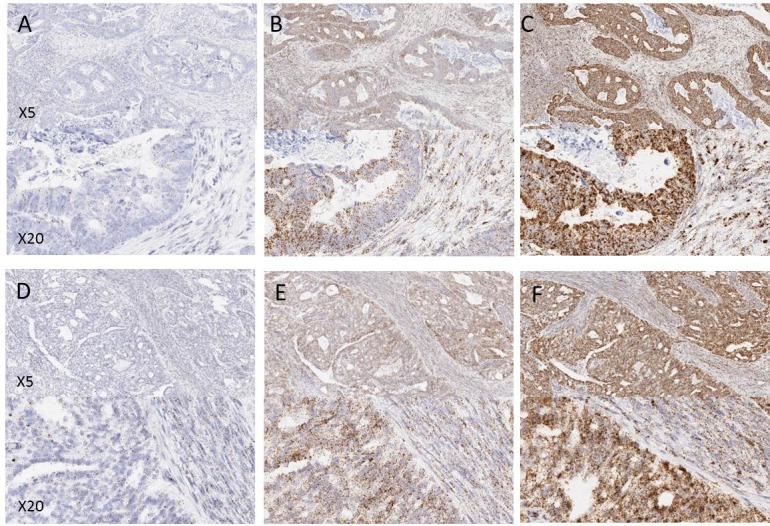
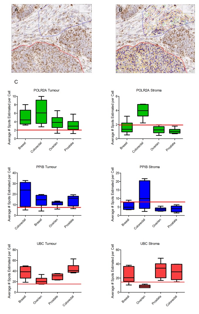
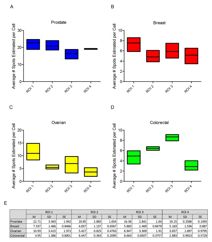
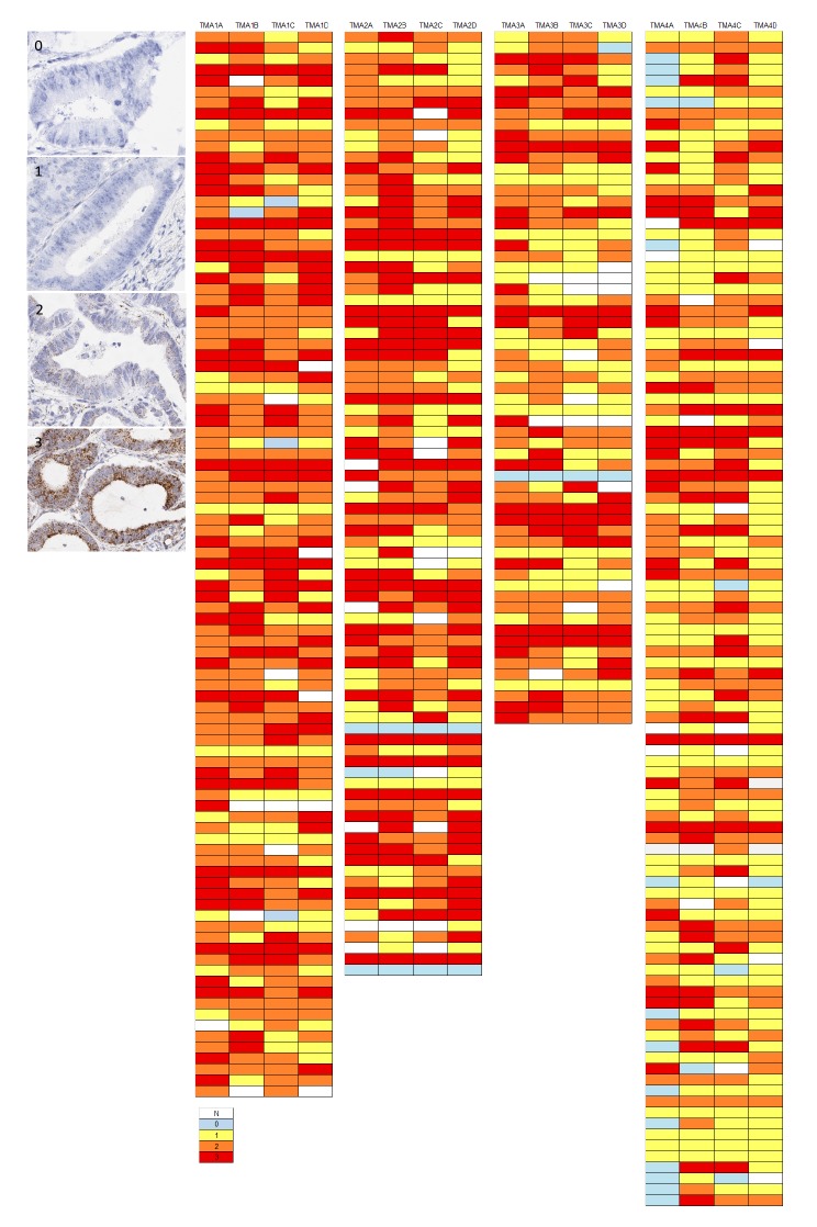
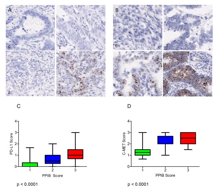
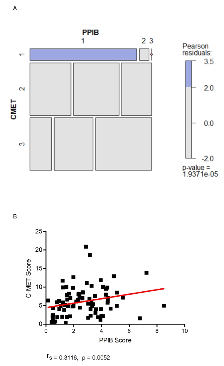
Immunohistochemistry remains the overwhelming technique of choice for test biomarker evaluation in both clinical or research settings when using formalin-fixed, paraffin embedded tissue sections. However, validations can be complex with significant issues about specificity, sensitivity and reproducibility. The vast array of commercially available antibodies from many vendors may also lead to non-standard approaches which are difficult to cross-reference. In contrast mRNA detection, by in situ hybridization (ISH) with sequence specific probes, offers a realistic alternative, with less validation steps and more stringent and reproducible assessment criteria. In the present study mRNA ISH was evaluated in prospectively and retrospectively collected FFPE samples within a cancer biobank setting. Three positive control probes, POLR2A, PPIB and UBC were applied to FFPE sections from a range of tumour types in FFPE whole-face (prospective collection) or TMA (retrospective collection) formats and evaluated semi-quantitatively and by image analysis. Results indicate that mRNA can be robustly evaluated by ISH in prospectively and retrospectively collected tissue samples. Furthermore, for 2 important test biomarkers, PD-L1 and c-MET, we show that mRNA ISH is a technology that can be applied with confidence in the majority of tissue samples because there are quantifiable levels of control probes indicating overall mRNA integrity.

Keywords: FFPE; Integrity; Pathology Section; in situ hybridization; mRNA.


Conflict of interest statement

CONFLICTS OF INTEREST The authors declare no conflicts of interest.

Figures

Figure 1
Figure 1. Examples of control probe expression in FFPE tissues from a prospectively collected cohort of samples
(A and D) POLR2A, (B and E) PPIB, (C and F) UBC. A, B and C are examples from colorectal tissue, D, E and F are examples from ovarian tissue. Note the increasing expression level from POLR2A to UBC.
Figure 2
Figure 2. Quantitative expression by Spotstudio image analysis of three control probes in 6 cancer samples from 4 different tumour types
(A). Region of tumour and stroma in a colorectal cancer case showing high expression of PPIB. (B). Example of the ROI selections for spot studio analysis in both tumour (red) and stroma (blue) compartments. Note the increased expression of PPIB in the tumour cells compared to stroma. (C) Graphed results are presented as average # spots estimated per cell in both tumour and stromal compartments. In all cases the horizontal red line depicts the lower threshold of expected expression. In almost all tumour compartments the expression of control probes would determine the tissues as fit-for-purpose for test biomarker analysis. Cut offs in stromal compartments are less robust.
Figure 3
Figure 3. Boxplots depicting the average number of spots per cell of PPIB at three levels in four regions of interest (ROI)
Prostate (A), breast (B), ovarian (C) and colorectal cancer (D). While variations are observed all ROIs irrespective of depth into the block would be suitable for test probe analysis. Table of descriptive statistics (E) summarising the mean, standard deviation and standard error between each level for the four ROIs.
Figure 4
Figure 4. Examples of variation in PPIB expression (0-3) in individual TMA cores in a CRC series
The accompanying illustration depicts the range of expression in 4 TMAs. Each TMA is represented by 4 replicate cores from the same donor FFPE block – 1A, 1B, 1C & 1D.
Figure 5
Figure 5. PPIB expression in TMA cores in a CRC series by block age of the donor FFPE sample
Almost as many cases have moderate or high levels of expression in 2004 as in 2008. The rate of negative cores is actually higher in 2008 but still very low as a percentage of total cases. Green – PPIB score of 0; Blue – PPIB score of 1; Red – PPIB score of 2/3.
Figure 6
Figure 6
(A) Varying levels of PD-L1 expression (0-3) in CRC TMA cores. (B) Varying levels of MET expression (0-3) the CRC TMA cores. Boxplots depicting the manual scores for PD-L1 (C) and C-MET (D) compared to manual PPIB scores on TMA cores from 40 cases. A Kruskal-Wallis Test was performed to assess differences in test probe mean score versus cases with PPIB categorical scores 1-3. A p value of less than 0.05 was considered statistically significant.
Figure 7
Figure 7
Mosaic plot (A) demonstrating individual contributions of categorical PPIB and c-MET scores with Pearson residual base shading. A p value of less than 0.05 was considered significant. Correlation plot (B) demonstrating the relationship of digital scores generated for c-MET to PPIB when a manual PPIB score of 1 was given. Correlation was calculated using Spearman’s Rank.

References

    1. Kunju LP, Carskadon S, Siddiqui J, Tomlins SA, Chinnaiyan AM, Palanisamy N. Novel RNA hybridization method for the in situ detection of ETV1, ETV4, and ETV5 gene fusions in prostate cancer. Appl Immunohistochem Mol Morphol. 2014;22:e32–40. - PMC - PubMed
    1. Baleriola J, Walker CA, Jean YY, Crary JF, Troy CM, Nagy PL, Hengst U. Axonally synthesized ATF4 transmits a neurodegenerative signal across brain regions. Cell. 2014;158:1159–72. - PMC - PubMed
    1. Schalper KA, Velcheti V, Carvajal D, Wimberly H, Brown J, Pusztai L, Rimm DL. In situ tumor PD-L1 mRNA expression is associated with increased TILs and better outcome in breast carcinomas. Clin Cancer Res. 2014;20:2773–82. - PubMed
    1. Lewis C, McQuaid S, Hamilton PW, Salto-Tellez M, McArt D, James JA. Building a ‘Repository of Science’: the importance of integrating biobanks within molecular pathology programmes. Eur J Cancer. 2016;67:191–99. - PubMed
    1. Bingham V, Ong CW, James J, Maxwell P, Waugh D, Salto-Tellez M, McQuaid S. PTEN mRNA detection by chromogenic, RNA in situ technologies: a reliable alternative to PTEN immunohistochemistry. Hum Pathol. 2016;47:95–103. - PubMed