Clinical features of visual disturbances secondary to isolated sphenoid sinus inflammatory diseases
- PMID: 29212484
- PMCID: PMC5717843
- DOI: 10.1186/s12886-017-0634-9
Clinical features of visual disturbances secondary to isolated sphenoid sinus inflammatory diseases
Abstract
Background: Visual disturbances associated with isolated sphenoid sinus inflammatory diseases (ISSIDs) are easily misdiagnosed due to the nonspecific symptoms and undetectable anatomical location. The main objective of this retrospective case series is to investigate the clinical features of visual disturbances secondary to ISSIDs.
Methods: Clinical data of 23 patients with unilateral or bilateral visual disturbances secondary to ISSIDs from 2004 to 2014 with new symptoms were collected. Collected data including symptoms, signs, neuroimaging and pathologic diagnosis were analyzed.
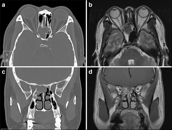
Results: There were 14 males and 9 females, and their ages ranged from 31 to 83 years. Fifteen patients suffered blurred vision and 11 patients suffered binocular double vision, including 3 patients who had unilateral visual changes and diplopia simultaneously. Headache was observed in 18 patients, and orbit pain/ocular pain in 8 patients. Other presenting symptoms included ptosis (4 patients) and proptosis (1 patient). Only 5 patients had nasal complaints. The corrected visual acuities were between NLP to 20/20. Patients with diplopia included 5 with unilateral oculomotor nerve palsy and 6 with unilateral abducens nerve palsy. All patients performed orbital/sinus/brain radiologic examination and found responsible lesions in sphenoid sinus. All patients underwent endoscopic sinus surgery, and 9 patients were found to suffer sphenoid mucocele, 9 with fungal sinusitis, and 5 with sphenoid sinusitis. Visual disturbances improved in 6 patients, and all the patients with diplopia had a postoperative recovery.
Conclusion: Visual disturbances resulting from ISSIDs are relatively uncommon, but it is crucial that the patient with new vision loss or diplopia and persistent headache or orbit pain be evaluated for the possibility of ISSIDs especially before corticosteroid administration.
Keywords: Diplopia; Headache; Isolated sphenoid sinus inflammatory diseases; Visual disturbance.
Conflict of interest statement
Ethics approval and consent to participate
The study was approved by the Institutional Review Board and Ethics Committee of Capital Medical University.
Consent for publication
Written informed consent was obtained from all patients to publish their cases in this case series.
Competing interests
The authors declare that they have no competing interests.
Publisher’s Note
Springer Nature remains neutral with regard to jurisdictional claims in published maps and institutional affiliations.
Figures


References
-
- Lin Y, Fang S, Ho H. Isolated sphenoid sinus disease: analysis of 11 cases. Tzu Chi Medical Journal. 2009;3:227–232. doi: 10.1016/S1016-3190(09)60044-6. - DOI
MeSH terms
LinkOut - more resources
Full Text Sources
Other Literature Sources
Medical

